INFLAMMATION PART II BY PRO.DR.SOHEIR SAAD PowerPoint PPT Presentation
Title: INFLAMMATION PART II BY PRO.DR.SOHEIR SAAD
1
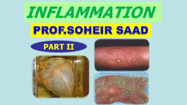
INFLAMMATION
PROF.SOHEIR SAAD
PART II
2
Non suppurative inflammation Includes
- Catarrhal inflammation.
- (2) Serous inflammation
- (3) Sero-fibrinous inflammation.
- (4) Fibrinous inflammation.
- (5) Membranous inflammation.
- (6) Haemorrhagic inflammation.
- (7) Necrotizing inflammation.
- (8) Allergic inflammation.
3
NON-SUPPURATIVE INFLAMMATION
- Cattarhal inflammation
- (e.g. rhinitis, gastritis, colitis.)
- When mild inflammation of the mucous membrane
stimulate excess mucous secretion, the exudate is
thick and viscid. - When secondary pyogenic infection supervenes,
it becomes mucopurulent inflammation. - The exudate contains mucous and pus
4
(1)Cattarhal inflammation
(e.g. rhinitis, gastritis, colitis.)
- When mild inflammation of the mucous membrane
stimulate excess mucous secretion, the exudate is
thick and viscid. - When secondary pyogenic infection supervenes, it
becomes mucopurulent inflammation. - The exudate contains mucous and pus
5
(No Transcript)
6
Produces thin serous exudate with low fibrin
content e.g. blisters of superficial burns.
2. Serous inflammation
By increasing the content of fibrin in the
exudate, you get serofibrinous inflammation
This is a feature of inflammation of serous and
synovial membranes e.g. pericarditis, pleurisy,
arthritis.
4.serofibrinous inflammation
7
(No Transcript)
8
(No Transcript)
9
(No Transcript)
10
(No Transcript)
11
(No Transcript)
12
Still with more fibrin in the exudate, It
results fibrinous inflammation, the best
example for this type is lobar pneumonia. This is
because the invading organisms are virulent
producing severe injury to the vascular
endothelium thus allow more leakage of the large
fibrin molecule.
4.fibrinous inflammation
5. Haemorrhagic
The presence of large number of red cells in the
exudate indicates severe damage to the
blood vessel, so allows more diapedesis of
RBC, and the inflammation is called Haemorrhagic
e.g. plague, and anthrax.
13
Anthrax infection in humans presents as a
boil-like skin lesion that eventually forms an
ulcer with a black center (eschar). The black
eschar often shows up as a large,
14
6. Pseudomembranous inflammation
- When there is severe damage to the mucous
membrane caused by the exotoxins liberated from
the invading organism like the diphtheria
bacillus, or the chigella chiga (causing
bacillary dysentery) - The desquamated epithelium become mixed with the
fibrinous inflammatory exudate to form a
pseudomembrane.
15
(No Transcript)
16
(No Transcript)
17
- Occurs whenever the inflammation is severe
resulting in thrombosis of the
veins in the area, leading to ischaemic necrosis.
- This may be followed by putrefaction and end with
gangrene e.g. gangrenous appendicitis.
Gangrenous necrotizing inflammation
The fluid is usually serous or
serofibrinous, the cellular component is
characterized by an excess of eosinophils e.g.
hypersensitivity reactions (asthma, urticaria).
8. Allergic inflammation
18
Non-suppurative inflammation
Name Occur in Characterized by
Catarrhal Mild inflammation in mucous membrane of respiratory or alimentary tracts e.g. common cold and catarrhal appendicitis Exudates rich in mucous
Serous Mild inflammation in serous surface such as pleural cavity, joint cavity where no damage in endothelium ex. Tuberculosis pleurisy and Common blisters Extensive watery low protein exudates
Fibrinous Outpouring of exudates with high protein and less volume ex. in lobar pneumonia due to Streptococcus pneumonia pericardium inflammation Exudates rich in fibrinogen
Membranous Fibrinous inflammation in which network of fibrin entangling inflammatory cells and bacteria forms pseudo-membrane. Example Diphtheria , Bacillary dysentery. Yellowish grey pseudo membrane rich in fibrin , polymorphs necrotic tissues
Hemorrhagic In blood vessels e.g. in plague Exudates rich RBCs
Gangrenous Acute appendicitis Necrotic tissues resulting from thrombi or emboli
Allergic Result to Ag Ab reaction Hypersensitivity Presence of edema increase in vascularity.
19
Chemical Mediators
20
The Inflammatory Chemical Mediators
- The inflammatory response is precipitated by
injury, but it is mediated by chemicals derived
from plasma, cells or damaged tissue. - The chemical mediators of inflammation are
usually present within the body in an inactive
form that is activated by injury.
21
The Inflammatory Chemical Mediators
- A) Permeability factors
- 1. Amines.
2. Kinin.
3. Activated fractions. (C3a, C5a
compliment)
4. Prostaglandin.
22
B) Chemotactic Factors
(Leucotactic factors)
- 1. Compliment related factors.
- 2. Bacterial factors.
3. Protein stored in PNL granules.
4. Tissue factors.
5. Eosinophilic chemotactic factor of
anaphylaxis.
6. Lymphokines.
23
A) Permeability factors These produce
vasodilatation and increased vascular
permeability.
- 1. Amines
- Particularly histamine and serotonin (5
hydroxytrpytamine) They are stored in granules of
mast cells, basophils and platelets. - They are responsible for the early phase of
acute inflammation (30-90 min).
- 2. Kinin
- They are normally circulating in plasma in an
inactive form and become activated by the
presence of antigen antibody complexes, by
bacterial endotoxin and by polymorph lysosomal
enzymes. - The most important of this group is bradykinin.
- Their action is more prolonged, up to 2½ hours.
- They also cause pain
24
Activated fractions (compliment)
- They are potent but their action is short lived.
- They cause release of histamine from mast cells.
- C3 component increases vascular permeability.
- C5 component increases vascular permeability
(more potent than C3). It is also chemotactic for
neutrophils macrophages - C5,6,7 complex is chemotactic for neutrophils
macrophages. It has no permeability effect.
25
- It is a long-chain fatty acid present in
almost all cells, they are rapidly catabolised
and are not stored in the body. - Prostaglandin E1 and E2 are thought to be
important in sustaining the later phases of the
inflammatory response (3-24 hours). - They act in part by liberating histamine.
- It also causes pain.
- It is interesting to know that action of
aspirin and allied drugs as anti-inflammatory,
antipyretic and analgesic is through inhibiting
the synthesis of prostaglandin from the injured
tissue.
4. Prostaglandin
26
- (B) Chemotactic (Leucotactic factors)
- They direct the movement of phagocytic cells
towards the irritant - Compliment related factors
- (i) Cleavage fragments of C3 and C5, similar but
not identical with C3a and C5a (anaphylatoxins). - (ii) C567, an activated component, is chemotactic
for PNL. - 2. Bacterial factors
- (i) Soluble low molecular weight products are
directly chemotactic. - (ii) Bacterial proteases activate the compliment
system and cleave C3 and C5. - 3. Protein stored in PNL granulesHave both
vasoactive and chemotactic activity.
27
4. Tissue factors Tissue injury results in
release of a C3 cleaving enzyme from the damaged
cells. 5. Eosinophilic chemotactic factor of
anaphylaxis Released from sensitized mast cells
on exposure to the specific antigen, e.g. atopic
reaction such as urticaria, hay fever and
asthma. 6. Lymphokines Released by sensitized
lymphocytes in delayed hypersensitivity reactions
is chemotactic for macrophage.
28
(No Transcript)
29
FATE AND OUTCOME OF ACUTE INFLAMMATION
1. Resolution
2. Healing
3. Spread
4. Chronic inflammation
5. Death
30
1. Resolution
- This means complete return of the tissue to
normal. - It occurs with mild inflammation provided
that there - has been NO tissue destruction.
- The inflammatory exudate reabsorbed and the
tissues restored to normal, e.g. uncomplicated
lobar pneumonia.
2. Healing
When the tissues have been destroyed, healing
occur by either repair or regeneration.
31
- Direct Spread through tissue spaces e.g.
cellulitis. - (ii) Lymphatic Spread Through producing
lymphangitis and lymphadenitis. - (iii) Blood spread producing Bacteraemia,
Toxemia, Septicemia and Pyaemia.
3. Spread
32
- Occurs when the organisms are not
- completely destroyed by the defensive
- mechanisms of the host.
- They remain in the body and provoke chronic
inflammatory and immunologic reactions. - From toxaemia or involvement of vital organs e.g.
heart (myocarditis), the brain
4. Chronic inflammation
5. Death
33
Outcome of Acute inflammation
spread
Death
34
(No Transcript)
35
CHRONIC INFLAMMATION
36
Chronic INFLAMMATION
37
Chronic Inflammation
Chronic Non-Specific
Chronic Specific
- chronic abscess
- chronic sinus
- chronic fistula,
- chronic ulcer.
GRANULOMA
NON Infective Sarcoidosis Forgein body
Infective e.g T.B ,S,B
38
- Chronic inflammation occurs when the body cannot
get rid of the irritant and remains in the body
in rather a state of equilibrium with the bodys
defensive mechanisms - It is a prolonged process in which tissue
destruction and inflammation are proceeding at
the same time as attempts at repair i.e.. - 1.Local tissue damage.
- 2. Inflammation response.
- 3. Attempts at healing and repair.
Chronic inflammation
Definition of chronic inflammation
39
Causes of chronic inflammation
- (1) Persistence of the agent, which caused the
acute inflammatory response, e.g. unevacuated
abscess. - (2) Insoluble irritant, foreign bodies such as
silica particles, talc powder, asbestos, catgut
suture etc. - (3) Poor local circulation as associated with
varicose veins of leg. - (4) Immunologic injury such as Rheumatoid
arthritis, Crohns disease. - (5) Resistant microorganisms such as T.B., Lepra
bacilli.
40
General characteristics of chronic inflammation
- Although the histologic appearance vary according
to the causative factor, yet they all share in
the following - The reaction is more proliferative (productive)
than exudative exudation of fluid - (2) The cellular reaction is a pleomorphic one,
consisting mainly of macrophage, lymphocytes and
plasma cells and sometimes eosinophils, PNL are
found in considerable number ONLY in chronic
suppurative lesions, such as chronic abscess,
chronic osteomyelitis. - (3) The arterioles show thick walls and narrow
lumens (endarteritis obliterans) in contrast to
the changes seen in acute inflammation. - (4) Fibrosis is a common feature, preceded by
granulation tissue. - (5) The chemical inflammatory mediators are
mainly the lymphokines.
41
- In Case of Non-Specific Chronic Inflammation
- The microscopic appearance gives no indication of
the cause. - Follows acute inflammation.
- The inflammatory cells are present in a diffuse
manner with more concentration around blood
vessels (perivascular infiltration), e.g. chronic
abscess, chronic sinus, chronic fistula, chronic
ulcer.
42
Chronic Specific Inflammation (Granulomatous
Inflammation)
- It has distinctive histologic appearance
characteristic of its causal agent, so in most
cases, it is easy to name the exact causative
irritant e.g. T.B., Bilharziasis. - Usually chronic from the start.
- Usually characterized by granuloma formation
and so called chronic granulomatous inflammation.
43
Granuloma
- Is defined as collection of chronic
inflammatory cells composed largely of
macrophage, starts as tiny granules, then
coalesce to form a mass hence the suffix oma
(tumour). - Are caused by insoluble particles that induce
cell mediated immune response. - The macrophages engulf the foreign material
and present them to T-lymphocytes which become
activated to which secrete cytokines, such as
IL-2 which activate other lymphocytes and IFN-d
which transform macrophages into epithelioid
cells and giant cells.
Granulomas
44
Activated lymphocytes MQ influence each other
release inflammatory mediators that affects other
cells.
45
(No Transcript)
46
(No Transcript)
47
(No Transcript)
48
- Classification of Granuloma-
- Infective caused by
- Bacteria, T.B., leprosy, syphilis, actinomycosis,
rhino-scleroma. - b) Parasites e.g. Bilharzia, Leishmania.
- c) Fungi and other microorganism e.g. Madura
foot. - 2. Non-infective Foreign body granuloma
- Usually caused by foreign body.
- Endogenous like fat (fat necrosis), cholesterol
crystal. - b) Exogenous like silica, asbestos, talc powder,
insoluble ligatures. - c) Granuloma of immunologic origin like Aschoff
body (rheumatic carditis), Crohns disease.
49
- Role of Inflammatory Cells in Inflammation
- Phagocytic cells
- Polymorphs
- Engulf bacteria and to a lesser extent
products of necrotic tissue, - redominate in the acute inflammatory
reaction. - Macrophage predominate in chronic granulomatous
inflammation, he macrophage is derived from
blood monocytes, tissue histiocytes,. - Giant cells They are formed either from fusion
of many macrophage or by repeated amitotic
division of one histiocyte. They clean and clear
the area for repair process. They are responsible
for the so called demolition phase.
- Types of Giant Cells
- Foreign body giant cells seen in cases of foreign
body granuloma. - 2. Langhans giant cells in T.B.
- 3. Aschoff cells of Rheumatic fever.
- 4. Tumour giant cells e.g. Reed-Sternberg cell in
Hodgkins disease and giant osteoclast in bone
tumours
50
- 2. Cells responsible for the immune reaction
- Lymphocytes
- B-cells for humeral immunity(undergo
transformation into - plasma cell (antibody
producing cell). - T-cells (CD4 CD8 ) for the cell-mediated
immune response - NK). Under antigenic stimulation, the
B-lymphocytes - Mast cells which are responsible for the
secretion of chemical mediators early in the
inflammatory lesions. - Eosinophils seen in Allergic and Helminthic
infection. They also secrete eosinophilic
chemotactic factor of anaphylaxis. - Fibroblast They have a major role in the
process of repair.
51
Name Neutrophil Eosinophil Basophil Monocyte Lymphocyte Plasma cell
Microphage Acidophile Basophil Macrophage Histocytes lymphocyte Plasmacyte
Shape Pale pink to blue, Minimal granulation. Red with eosin, Coarse granulation. Blue with eosin, Coarse granulation 1.5 to 2 times larger, Abundant fine granulation Agranular non-granulated, Large round nucleus Basophilic, Encentric nucleus
of WBCs 60-70 1-2 (50 in allergy) 1 4-6 30 Found in tissue only
Function Phagositic 1st defense Unknown but could neutralize histamine, serotonin and other kinins Unknown but contain histamine heparin Phagocytic 2nd defense element engulf bacteria, dead cells, debris dead neutrophils (pus cells) Antibodies production Late stage of the inflammation Primary source of specific Antibodies
Monocytes
Plasma cell
Plasma cell
52
Chronic Inflammation
Acute Inflammation
Presence of lymphocytes
- Is rapid in onset
- (typically minutes)
- insidious in onset
- Short duration
- lasting for hours or a few days
- Longer duration
- Characterized by
- exudation of fluid
- plasma (edema)
- emigration of PNLs
- Characterized by
Macrophageproliferation of bl.v. fibrosis
tissue necrosis.
53
Comparison between acute and chronic
inflammation
chronic acute
Delayed for months or years Immediate for few days Onset Duration
Persistent infections by microorganisms , Immune-mediated inflammatory diseases,Prolonged exposure to potentially toxic agents Infections Tissue necrosis from any cause, Etiology
The reaction is more proliferative (productive) than exudative predominantly cells lymphocytes Macrophages plasma cells fibroblasts vascular changes, edema, and predominantly neutrophilic infiltration,,and Macrophages Tissue reaction
lymphokines Vasoactive amines mediators
Tissue destruction with attempts for healing and fibrosis. Resolusion,healing,abscess,chronic inflammation outcome
54
(No Transcript)

