Medical Image Analysis PowerPoint PPT Presentation
1 / 18
Title: Medical Image Analysis
1
Medical Image Analysis
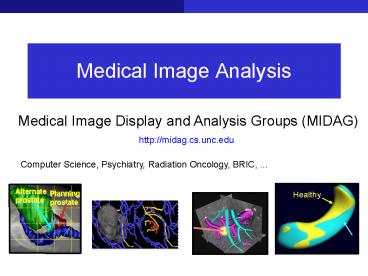
Medical Image Display and Analysis Groups (MIDAG)
http//midag.cs.unc.edu
Computer Science, Psychiatry, Radiation Oncology,
BRIC, ...
2
What is Medical Imaging?
Novel imaging methods have revolutionized
medicine.
...
1895 X-rays W. K. Rontgen
1960's Computed tomography Cormack and Hounsfield
1970's Magnetic Resonance Imaging Mansfield and
Lauterbur
Much of it happened throughout approx. the last
100 years.
Today, we can look into the human body in vivo.
3
Quantification of Image Information
Fluoroscope
1930s-1950s The frivolous shoe-store
Does my shoe fit?
Connection to computer science
Modern imaging methods produce increasing amounts
of data, which needs to be quantified
automatically.
4
Quantification of Image Information
How to reconstruct, visualize, and analyze huge
data quantities?
5
Small, smaller, even smaller ...
MRI
microscopy
electron tomography
organelles
(macro)molecules
tissue
cells
100 µm
10 µm
1 µm
1 mm
100 nm
10 nm
1 nm
0.1 nm
(µ)MRI/(µ)CT/PET
Light microscopy
Electron tomography
Electron crystallography
X-ray crytstallography
Nuclear magnetic resonance
6
Interesting CS Projects with Impact
- Helping people and advancing science
- Cancer treatment planning and delivery
- Diagnosing degree of malignancy and associated
drug effects - Study of autism and other diseases of the brain
- Diagnosis of brain defects at birth for treatment
- Animal studies of disease effects
- Computer-assisted surgery
- Visualizations for diagnosis of joint injuries
- Study of cellular and subcellular phenomena
- ....
7
Tremendous Educational Breadth
- Areas of study involved
- Graphics
- Applied MathematicsGeometry, Numerics, Fluid
Flow, ... - Statistics
- Software design and algorithms
- User interfaces
- Medical imaging
- Exposure to the medical problem
8
Great Environment to Learn and Research
- Computer Labs
- CS Graphics Image Analysis Lab
- RadOnc Computational Radiation Treatment Lab
- Psychiatry Neuro Image Analysis Lab
- Surgery Computer-Assisted Surgery and Imaging
Lab - Radiology IDEA lab
- BRIC Biomedical Research Imaging Center
- Medical treatment research labs
- As above, Pediatrics, Neurology, Psychology,
Dentistry, Cell Developmental Biology,
Pharmacology, Dermatology, ...
9
Medical Image Analysis in the CS department
- Stephen Pizer, PhD, Kenan Prof. CS ( RadOnc,
Radiology, BME) Statistical geometry of
objects, medial descriptions. - Marc Niethammer, PhD, Ass. Prof. CS/BRIC
Shape analysis, advanced imaging modalities. - Martin Styner, PhD, Ass. Prof. Psych CS, Neuro
Image Analysis Lab Shape analysis, brain image
analysis. - Mark Foskey, PhD, Ass. Prof. RadOnc CS
Cancer therapy, geometric computation. - J. Stephen Marron, PhD, Amos Hawley Prof.
Stat/OR CS Statistical methods. - Julian Rosenman, MD, PhD, Prof. RadOnc
CS radiation treatment, visualization. - Dinggang Shen, PhD, Associate Prof.
Radiology/BRIC Image Registration and Analysis.
10
Biomedical Imaging Research Center (BRIC)
- To support biomedical imaging research
- Animal studies, inflammation, cancer, knockout
- Microscopic and nanoscopic images
- Cells, molecules, etc.
- Human studies of disease development
- Individual human diagnoses and therapies
11
Brain Imaging
Analysis of white matter connectivity.
Analysis of gray matter morphology.
Images from Grays Anatomy
White-matter fibers connect gray-matter
structures.
Quantify brain changes. How?
12
Structural and Diffusion Weighted (DW-) MRI
White matter bundles from DW-MRI the main
direction of water diffusion in the brain
Structural MRI
Fiber bundle directions
Tissue type GM/WM
Image Gordon Kindlmann
13
Normal Brain?
What is normal?
- Normal development
- Aging (Fitness)?
- Atlas generation
Brain during NCAA tournament
Normal
Schizophrenic
14
Project (RA) Longitudinal Brain Analysis
source
target
How does a brain develop?
Algorithm design real experiment
Structural and diffusion MRI
source
warped source
w/ Martin Styner and Brad Davis
15
Project (991) Knee Imaging
To aid in detection and to evaluate progression
of osteoarthritis.
Osteo bone Arthro Joint Itis inflammation
Bone surfaces are protected by cartilage, which
is lost or reduced in osteoarthritis causing pain.
How does cartilage thickness change over time?
Affects millions of people in the US.
16
Project (991) Electron Tomography
Image analysis for synapses, e.g., where are the
vesicles, where are cell membranes, what is the
architecture of the active zone?
3D image volume
Axon-gtDendrite
tilted views
analysis
reconstruction
w/ Russ Taylor and Alain Burette
17
Open Position and 991s
- RA position and a variety of 991 projects
available. - Longitudinal brain analysis.(MRI, structural and
diffusion)? - Knee cartilage changes over time. (structural
MRI)? - Image analysis for synapses (electron
tomography)? - Tracking of focal adhesions (microscopy), ...
.... challenging, interdisciplinary, driven by
medical research.
If interested, come see me between 6-8pm or any
time by appointment. Marc Niethammer office
SN 219 email mn_at_cs.unc.edu
18
... and finally ...
If you are interested in medical imaging in
general, sign up for
Comp 775 Introduction to Medical Image Analysis
- Offered this Fall
- Mon/Wed 200pm-315pm
- FB 007
I hope to see you all there ... -)?

