
DNA, chromatin and the nucleus PowerPoint PPT Presentation
1 / 20
Title: DNA, chromatin and the nucleus
1
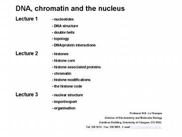
DNA, chromatin and the nucleus Lecture 1 -
nucleotides - DNA structure - double
helix - topology - DNA/protein
interactions Lecture 2 - histones - histone
core - histone associated proteins -
chromatin - histone modifications - the
histone code Lecture 3 - nuclear structure -
import/export - organisation Professor N.B. La
Thangue Division of Biochemistry and Molecular
Biology Davidson Building, University of Glasgow,
G12 8QQ. Tel 330 5514. Fax 330 5859. E-mail
N.LaThangue_at_bio.gla.ac.
2
(No Transcript)
3
- Information is Stored in the Code Letters of DNA
- All hereditary information is stored in genes,
which are - parts of giant DNA molecules
- Genes code for the amino acids of proteins
- DNA is the archival copy of the code- kept in
nucleus - where it is protected repaired
- DNA is organized with special proteins into
chromosomes - For protein synthesis a working copy of the
code is made - from RNA
- Overall scheme DNA -gt RNA -gt protein
4
- The Code is Based Upon the Structure of DNA
- DNA has a sugar-phosphate backbone- sugar is
deoxyribose - DNA also has 4 types of nucleotide base A, C,
G, T - A adenine C cytosine G guanine T
thymine - Molecule is a double helix 2 complementary
strands where A T, C G - The term"complementary" refers to the fitting
together of 2 molecules like - hand and glove
- In DNA complementary bases make good hydrogen
bonds with one another - Strands of helix are held together by hydrogen
bonds between the bases - This allows DNA to unwind for duplication and
transcription - (S sugar P phosphate B base)
-
5
Bases, Nucleosides, Nucleotides
The basic building blocks of nucleic acids are
the nucleotides. Each has three components
- A heterocyclic base
- There are two types of base
There are two purine bases commonly found in
nucleic acids
There are three pyrimidine bases commonly found
in nucleic acids
6
Nucleosides and Nucleotides
- A 5 carbon (pentose) sugar
- The sugars are in the furanose (ring) form and
can be - deoxyribose (in DNA)
- ribose (in RNA)
- The base is attached to the sugar by a glycosidic
bond at C1'.
- Phosphate moieties esterified to the C5' of the
sugar
7
The structure of B-DNA The practical
breakthrough in Watson Crick's search for the
structure of DNA
A base pair formed between a guanine nucleotide
and a cytosine nucleotide
A base pair formed between a thymine nucleotide
and an adenine nucleotide
8
- Features of the Watson-Crick model of B-DNA
- It is an anti-parallel double helix.
- It is a right-handed helix.
- The base-pairs are perpendicular to the axis of
the helix. - The axis of the helix passes through the centre
of the base pairs. - Each base pair is rotated by 36 degrees from the
adjacent base pair. - The base-pairs are stacked 0.34 nm apart from one
another. - The double helix repeats every 3.4 nm, i.e. the
pitch of the double helix is 3.4 nm. - B-DNA has two distinct grooves a MAJOR groove
and a MINOR groove.
9
The real structure of B-DNA
Watson and Crick's structure was a just a model
In 1980, Richard Dickerson and Horace Drew
solved the structure of
5'-CGCGAATTCGCG-3'
10
Energetics of B-DNA Three forces are responsible
for the stability of the B-DNA double helix
- Hydrophobic base-stacking interactions (van der
Waals forces) between adjacent base pairs. - Hydrogen bonds forming the base-pairs.
- Hydrogen bonds due to the formation of a water
spine in - the minor groove.
11
Variations in the structures of DNA
poly(A) tracts causes a bending of 18 degrees
palindromes, hairpins, pseudoknots cruciforms
A-DNA there is no water spine minor groove is
about as wide as the resulting major groove.
It is not clear if the A-DNA conformation exists
in vivo.
Z-DNA crystallizing the self-complementary
hexanucleotide, CGCGCG. This particular molecule
adopted a LEFT-HANDED double helix.
Z-DNA seems to form most readily in sequences
that alternate purines and pyrimidine
12
Supercoiling and Topoisomerases
- Supercoiling
- The total amount of DNA in any individual cell
seems very large - DNA can adopt a compact configuration due to
supercoiling. - Supercoiling is a physical rearrangement of the
DNA double helix that allows it to confirm more
closely to the ideal B-DNA structure. - Implications for transcription and replication
unwinding/unpairing must occur so that mRNA or
DNA copies can be made. - Linking Number and Superhelical Density
Supercoiling in circular DNA molecules is the
number of times the two phosphodiester backbones
wrap around one another in a given distance. - In bacteria, the superhelical density is 0.06.
- Topoisomerases supercoiling is carefully
controlled by the action of topoisomerases
Naturally occurring DNA is underwound.
13
Topoisomerases
There are two classes of topisomerase Type 1
topoisomerases remove supercoils through a
mechanism that involves breaking only one of the
two phosphodiester backbones.
The best-characterized member of this class in
E. coli is Topoisomerase I. 864 amino-acids in
length and is monomeric encoded by the topA gene.
- formation of a covalent intermediate between a
tyrosine residue and the - phosphodiester backbone.
- nucleophilic attack from the hydroxyl group of
tyrosine to a phosphorus atom - creates a phosphodiester link between the
enzyme and the DNA
14
- Type 2 topoisomerases
- Both phosphodiester backbone chains are broken
simultaneously. - E. coli, Topoisomerase II better known as DNA
Gyrase. - E. Coli DNA gyrase is a tetrameric protein
consisting of two A subunits (875 aas) and two B
subunits (804 aas). - Topoisomerases are essential enzymesmutations in
any of the genes coding for - topoisomerases are usually lethal. They are
therefore targets for antibiotics and - other drugs. Novobiocin, doxorubicin, etoposide.
15
Challenges of transcriptional control in a
mammalian cell
- 30,000 genes
- Genome size 3 x 10⁹ base pairs
- DNA organized into chromatin of varying levels of
compaction - Developmental requirements
- Homeostatic requirements
16
Transcriptional control in eukaryotic cells
- Transcription factors are regulated by
intracellular and extracellular signals
- Chromatin, sequence specific factors,
co-activators, co-repressors, and basal machinery
are all targets of signaling pathways
- Transcription factors work in a combinatorial
manner to achieve different transcriptional
outputs
17
Sequence-specific DNA-binding Transcription
Factors Are the Apex at the Interface of Genetic
Regulatory Information and Other Transcription
Regulators
18
(No Transcript)
19
(No Transcript)
20
Transcription factors act in a combinatorial
manner to regulate gene expression
Recommended
