ULTRASOUND - PowerPoint PPT Presentation
1 / 1
Title:
ULTRASOUND
Description:
... coupling the electrical circuitry onto the same chip as the transducer array. ... onto a single CMOS chip, an advance made possible by new transducer ... – PowerPoint PPT presentation
Number of Views:74
Avg rating:3.0/5.0
Title: ULTRASOUND
1
ULTRASOUND
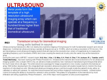
Metal posts form the template of a high
resolution ultrasound imaging array which can
operate at a frequency a hundred times higher
than that of traditional biomedical ultrasound.
J. Am. Ceram. Soc., in press (2006)
Penn State MRSEC, DMR-0213623
Transducer arrays for biomedical imaging living
cells bathed in sound
IRG3
Ultrasound array transducers offer
non-destructive real-time imaging of living
tissues for both fundamental research and
clinical diagnoses. Commercial ultrasound arrays
operate at frequencies up to 16 MHz, which
provides a resolution of 50 microns. We are
developing a novel ultrasound imaging system that
will improve the resolution two orders of
magnitude by increasing the operating frequency
up to hundreds of MHz and close-coupling the
electrical circuitry onto the same chip as the
transducer array. During the past year a MRSEC
research team (H.S. Kim, I. Kim, I. G. Mina, S.
K. Park, K. Choi, T. N. Jackson, R. L. Tutwiler,
and S. Trolier-McKinstry), has been working with
Phillips Research to miniaturize the transmit
and receive electronics for an ultrasound imaging
system onto a single CMOS chip, an advance made
possible by new transducer manufacturing
techniques that allow for much lower drive
volt-ages. The entire electronics package for
image acquisition is now closely coupled to the
transducer array. With further development, this
integrated high-resolution ultrasound system will
enable researchers to monitor the drug reactions
of individual live cells. Miniaturized ultrasound
imaging arrays could be brought inside the
patient's body, either on catheters or within
pill cameras, for real-time tissue biopsies and
sub-surface imaging in turbid environments.
Limited motion control (including the ability to
reorient the camera) is also possible. Tem-plates
for preparation of the transducer by mold
infiltration are being supplied by Philips
Research, one of the major world suppliers of
ultra-sound equipment.


