Lecture BIOD 4: Prokaryotic Cell Structure PowerPoint PPT Presentation
1 / 18
Title: Lecture BIOD 4: Prokaryotic Cell Structure
1
Lecture BIOD 4 Prokaryotic Cell Structure
Function
- Morphology of Bacteria
- Prokaryotic cells are generally much smaller
than Eukaryotic cells.
- Although similar in morphology, bacteria
remarkably vary in size, shape arrangement.
- Most bacteria come in one of three basic
shapes coccus, rod or bacillus, and spiral.
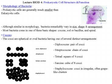
- Coccus
- The cocci are spherical or oval bacteria having
one of several distinct arrangements
- Diplococcus pair of cocci
- Streptococcus chain of cocci
- Tetrad square of 4 cocci
- Sarcina cube of 8 cocci
- Staphylococcus cocci in irregular, often
grape-like clusters
2
- Bacillus
- Bacilli are rod-shaped bacteria. They divide in
one plane producing one of the following
- Bacillus a single bacillus
- Coccobacillus oval and similar to a coccus
- Streptobacillus a chain of bacilli
- Spiral
- A helical or corkscrew-shaped bacterium.
Spirals come in 1 of 3 forms
- Vibrio curved or comma-shaped rod
- Spirillum thick, rigid spiral
- Spirochete thin, flexible spiral
3
- Pleomorphic
- Bacteria that are variable in shape lack a
single characteristic form.
- Prokaryotic Cell Organization
- Bacteria are prokaryotic, single-celled,
microscopic microorganisms
- Generally much smaller than eukaryotic cells
- Very complex despite their small size.
- Structurally, a typical bacterium usually
consists of
- a cytoplasmic membrane surrounded by a
peptidoglycan cell wall maybe an outer membrane
- a fluid cytoplasm containing a nuclear region
(nucleoid) and numerous ribosomes and
- often various external structures like a
glycocalyx, flagella, and pili.
4
- Nucleoid
- The most striking difference between
procaryotes and eucaryotes is the packaging of
the genetic material.
- Eucaryotes have 2 or more chromosomes contained
within a membrane-delimited organelle, the
nucleus.
- Procaryotes chromosome, single circle dsDNA, is
located in irregular shaped region called
nucleoid (also called nuclear body, chromatin
body, nuclear region).
- DNA (deoxyribonucleic acid) the nucleic acid
that constitutes the genetic material of all
cellular organisms. Polymer of nucleotides
connected via a phosphate-deoxyribose sugar
backbone.
5
- Many bacteria contain Plamids in addition to
their chromosome.
- Plasmids are circular dsDNA molecules,
replicate independently of chromosome and may
integrated in it not required for cell growth
and reproduction.
- Plasmids carry extra genes that render bacteria
drug-resistant, give them new metabolic
activities, make them pathogenic!!!!, etc.
- Cytoplasmic Matrix, Ribosome and Inclusion
Bodies
- Cytoplasmic Matrix is the substance lying
between the plasma membrane and the nucleoid
- It is a relatively featureless fluid about 70
water packed with Ribosomes and inclusion bodies.
- Inclusion Bodies are granules of organic or
inorganic material that are stockpiled for future
use (starch, fat, sulphur, gas vacuoles, etc.).
- Ribosomes structures made of protein and RNA
responsible for synthesis of cellular proteins.
- Mesosomes
- Invaginations of plasma membrane in the shape
of vesicles, tubules and lamellae.
- Play a role in cell wall formation during
division
- Involved in chromosome replication and
distribution to daughter cells.
6
- Plasma Membrane
- Membranes are absolute requirement for all
living organisms interact in a selective fashion
with environment.
- Plasma Membrane is made of a phospholipid
bilayer with integral and peripheral proteins
embedded.
- Imbedded in the membrane are proteins. Some
stick completely through the membrane, while some
are planted so that they only protrude through
one surface.
- These proteins are held in the membrane by
virtue of hydrophobic and hydrophilic regions on
the protein.
- Functions of PM
- Separates the cell from its environment.
- Serves as a selective permeable barrier.
- Transport systems used for nutrient
uptake,waste excretion, protein secretion.
7
- PM is the location of a variety of metabolic
processes such as respiration, photosynthesis,
lipid synthesis and cell wall constituents
synthesis.
- Contains special receptor molecules that may
help bacteria detect and respond to environmental
changes.
- Cell Wall
- One of the most important parts of the
prokaryotic cell.
- Give shape, protect from osmotic lysis, protect
from toxic substances.
- Cell Wall is made of peptidoglycan polymer of
peptides (typically 4 amino acids long,
cross-linked to other chains) and glycans (made
of alternating amino sugars).
- Sugars found in peptidoglycan
- - N-acetylglucosamine (NAG).
- - N-acetylmuramic acid (NAM).
8
- Gram Negative vs Gram Positive
- Christian Gram developed the Gram stain (1884)
bacteria divided in 2 major groups Gram()
Gram(-).
9
- Gram Positive
- Thick homogeneous peptidoglycan layer.
- Large amount of teichoic acids not in Gram(-).
- Inner plasma membrane.
10
- Gram Negative
- More complex than Gram positive.
- Thick outer membrane contains lipids,
lipopolysaccharides and lipoproteins.
- Thin peptidoglycan layer.
- Periplasmic space.
- Plasma membrane.
11
- Mechanism of Gram Staining
- Difference between Gram() Gram(-) is due to
the physical nature of their cell walls.
- Gram() becomes Gram(-) when cell wall removed.
- Peptidoglycan acts as a permeability barrier
preventing loss of crystal violet.
- Ethanol is thought to shrink pores of the thick
peptidoglycan ?dye-iodine complex retained ?
bacteria remain purple.
- Gram(-) have very thin peptidoglycan.
- Ethanol treatment extract enough lipid from
wall and make more porous ? purple crystal
violet-iodine complex is more readily removed.
- When counterstained with safranin ? Gram(-)
bacteria turn pink.
12
- Flagella and Motility
- Need to be stained to be viewed under bright
field microscopy.
- The ultrastructure consists of flagellin, hook
and basal body.
- Flagellin hollow rigid cylinder constructed of
a single protein.
- Hook short curved segment that links flagellin
to the basal body.
- Basal body Series of rings that drive the
flagella.
13
- Flagellar arrangements
1. Monotrichous a single flagellum, usually at
one pole. 2. Amphitrichous a single
flagellum at both ends of the organism. 3.
Lophotrichous two or more flagella at one or
both poles. 4. Peritrichous flagella over
the entire surface.
- Flagellar synthesis
- Complex process involving at least 20 to 30
genes, and 10 or more genes that encode proteins
for hook and basal body.
- Flagellar movement
- Bacterium moves when the filament (in the shape
of rigid helix) rotates.
- Flagella act like propellers on a boat.
- The E. coli motor rotates 270 rpm.
- Vibrio alginolyticus rotates 1100rpm.
14
(No Transcript)
15
- Capsules, Slime Layers S Layers
- Layers of material outside the cell wall, vary
in thickness and rigidity.
- Protects bacteria from phagocytosis, viral
infections, pH fluctuation, osmotic stress, ...
- Help bacteria to attach to surfaces of solid
objects in aquatic environment and tissue
surfaces in plants and animals.
- Pili Fimbriae
- Tubular protein (short hair-like) structures
extending from a bacterial surface used for
attachment to environmental surfaces or cells.
- Sex pili attach to surface of other bacteria
during sexual mating.
16
- Chemotaxis
- Bacteria are attracted nutrients such as sugars
and amino acids.
- Taxis is a motile response to an environmental
stimulus.
- Chemotaxis is a response to a chemical gradient
of attractant or a repellent molecules in the
bacterium's environment.
- Chemotaxis is regulated by chemoreceptors
located in the cytoplasmic membrane or periplasm
of the bacterium bind chemical attractants or
repellents.
- Bacterial Endospore
- An endospore is not a reproductive structure
but rather a resistant, dormant survival form of
the organism, enable bacteria to resist harsh
environment.
- Endospores are quite resistant to high
temperatures (including boiling), most
disinfectants, low energy radiation, drying, etc.
- The endospore can survive possibly thousands of
years until a variety of environmental stimuli
trigger germination, allowing outgrowth of a
single vegetative bacterium.
- Spore formation follows a very complex
multistage process.
17
(No Transcript)
18
- Under conditions of starvation, especially the
lack of carbon and nitrogen sources, a single
endospores form within some of the bacteria. The
process is called sporulation.
- Transformation of dormant spores into active
vegetative cells is done in 3 stages - (1) Activation. (2)Germination. (3) Outgrowth.

