Pr PowerPoint PPT Presentation
1 / 1
Title: Pr
1
Role of the spleen in leukemogenesis induced by
bovine leukemia virus
Florins A.1,
Gillet N.1, Jean G.1, Thewis A.1, Schwartz-Cornil
I.2, Bonneau M.2, Hay J.B.3, Kettmann R.1,
Willems L1. 1Molecular and Cellular Biology,
FUSAGx, Gembloux, Belgium 2INRA, Jouy-en-Josas,
France 3University of Toronto, Toronto, Canada
are members of the FNRS.
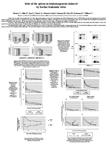
In this work, we aimed at characterizing the role
of the spleen during infection of sheep by bovine
leukemia virus (BLV). Experiments based on CFSE
labelling showed that B lymphocytes from infected
animals disappear faster from the blood compared
to the controls. This difference in dynamics
seemed to be mainly due to the B-CD11b
subpopulation. These cells are excluded from the
lymphatic compartment and migrate preferentially
towards the spleen. We thus hypothesized that
the spleen plays a major role in the kinetics of
B lymphocytes within BLV-infected sheep. In order
to study the involvement of the spleen, we
analyzed the percentages of various cellular
populations (B, B-CD11b, and cells expressing the
p24 viral antigen after brief culture) as well as
the proviral loads in the splenic artery and vein
of 2 infected sheep. No difference was observed
amongst all measured parameters. We next
performed a kinetic analysis of the B cell
population based on the use of intravenous CFSE
injection within 4 splenectomized sheep. We
observed that the difference in kinetics between
infected animals and the controls disappeared
after splenectomy. This report thus enlighten a
key role exerted by the spleen in the B cell
turnover of BLV-infected animals.
Figure 2 Dot plots obtained by flow cytometry
after PBMC labelling, 5 days after CFSE injection
(sheep 3002, infected by BLV). Labelling of the
p24 viral protein was performed after 16 hours of
culture in complete RPMI medium supplemented with
PMA/Ionomycin
Figure 1 Study of the PBMCs from jugular vein,
splenic artery and splenic vein of two control
sheep (4533 and 4534) and two infected animals
(4535 and 2672). (A) Percentages of B lymphocytes
within the PBMCs population. (B) Percentages of
B/CD11b in the B lymphocyte population. (C)
Percentages of PBMCs expressing p24 viral antigen
after 16 hours of culture in complete RPMI medium
supplemented with PMA/Ionomycin. (D) Average
numbers of viral copies per cell.
Figure 4 Percentages of CFSE positive cells in
the B lymphocyte population of infected sheep
(4535, 4536 and 3002) and controls (4533, 4534,
3004). (A) Short term, non splenectomized. (B)
Short term, splenectomized. (C) Long term, non
splenectomized. (D) Long term, splenectomized.
Figure 3 Percentages of CFSE-positive cells in
the T lymphocyte population of four
splenectomized sheep (two infected and two
controls). (A) T/CD4 lymphocytes. (B) T/CD8
lymphocytes. (C) T/?d lymphocytes.
Non splenectomized
Splenectomized
Figure 5 Percentages of CFSE-positive cells in
the B lymphocyte population within the jugular
and splenic veins (sheep 4544).
Figure 6 Percentages of CFSE-positive cells in
the B/CD11b- lymphocyte population for (A) non
splenectomized and (B) splenectomized sheep and
among B/CD11b lymphocytes for (C) non
splenectomized and (D) splenectomized sheep.

