Immunostaining of cells and tissues - PowerPoint PPT Presentation
1 / 31
Title:
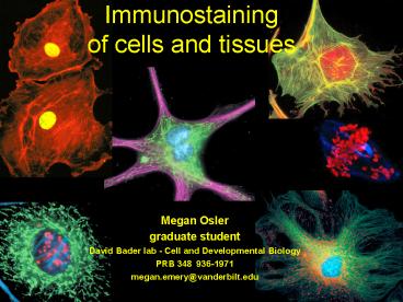
Immunostaining of cells and tissues
Description:
Methods of processing and detection. Variations. Purpose of Immunostaining. To ... donkey anti-goat. Y. Y. goat -mouse. Fc. How does the 2 recognize the 1 ... – PowerPoint PPT presentation
Number of Views:588
Avg rating:3.0/5.0
Title: Immunostaining of cells and tissues
1
Immunostaining of cells and tissues
- Megan Osler
- graduate student
- David Bader lab - Cell and Developmental Biology
- PRB 348 936-1971
- megan.emery_at_vanderbilt.edu
2
Objectives
- Purpose of technique
- Scientific basis
- Methods of processing and detection
- Variations
3
Purpose of Immunostaining
- To answer the question
- Where is my protein (antigen) located
- in a certain cell type or tissue?
Mouse gut Blue- goblet cells Red- actin in brush
border Green- stains nuclei
Endothelial cell Blue- tubulin Red-
F-actin Green- endosomes
4
What tool can I use to identify my
protein? (hint what immunoreagent recognizes
circulating antigens in your body?)
Antibodies
5
Antibodies specifically recognize a protein
epitope
Y
- Use an antibody raised to your protein and use it
as a marker to search for the antigen in
tissues/cells.
6
Generating (monoclonal) antibodies
Starting product Some or all of your protein
N
C
(you may be interested in this region)
7
How do you go from to ???
Y
Y
Y
8
Method (most common)
- Indirect Immunofluorescence
- Double antibody technique for signal amplification
9
Method (most common)
- Indirect Immunofluorescence
- Double antibody technique for signal amplification
10
How does the 2? recognize the 1??
11
2? binds the Fc portion of 1?
How does the 2? recognize the 1??
Y
Y
Fc
Very important You must match the species!
12
Method (most common)
- Indirect Immunofluorescence
- Multiple labeling
13
Y
Y
Y
Y
Y
Y
Y
Y
Y
Why double label?
14
Procedure overview
- Prepare sample
- Fixation/Permeabilization
- Blocking
- Primary antibody incubation
- Secondary antibody incubation
- Prepare for viewing
- variations
15
Cell/Tissue preparation
- Cells
- grow on cover slips or
- in chamber slides
16
Fixation Purpose to preserve the cells/tissue
and to immobilize the antigen
GOOD
fix
- Common fixatives
- PFA (1-4)
- Alcohol (MeOH or EtOH)
- Histochoice (commercial)
- How do you choose which fix to use?
- Antibody dependent
live cell
17
Permeabilization Purpose to punch holes in
the cell membrane so antibodies can diffuse in to
bind the target protein
Y
Y
Y
Perm.
- Permeabilizing agents (cells only)
- Alcohols (also a fixative)
- Detergents (Triton-X, Tween)
Antibodies can bind intracellular proteins
18
BlockingPurpose to eliminate non-specific
binding of antibodies
Reagent (most common) 2 Bovine Serum Albumin
(BSA)
Y
Y
Sticky proteins bind the BSA, so your antibody
will only bind YOUR PROTEIN
BSA
19
Antibody incubationPurpose bind your protein,
amplify signal
Cells blocked with BSA
Wash with PBS in between steps and Perform in a
humid chamber to prevent drying out the slide
20
Prepare for viewing Mount slide
- Purpose protect slide add anti-fade reagent
- Place coverslip on slides with mounting reagent
(Polyaquamount) - Keep in the dark until viewing
21
Important controls
- Pre-immune serum/2? Ab control
- Why? To determine that labeling is not background
from the 2? Ab - Ab that you KNOW will work (actin)
- Why? To verify that your procedure worked
- Dilution series
- Why? To optimize the concentration of the
antibodies for the best possible signal
22
Immunofluorescence
- Pros
- Easy
- Fast- 1 day
- High contrast
- Confocal
- Attractive images
- Cons
- Fluorescent microscope
- Bleaching
- Not permanent
23
One more thing
- Not every fluorescent signal originates from an
antibody. - There are STAINS also.
- Uses? Cytotoxicity, apoptosis
DAPI- nuclear stain
MitoTracker- mitochondria
Phalloidin- F-actin
24
Tricks for success
- PAP pen
- Nail polish
- VWR marker
- Q-tips
25
Potential Issues
Signal Intensity antibody avidity to protein 2?
Ab binding to 1? Ab
Y
Y
- Epitope masking
Y
26
Variations
- Immunohistochemistry (enzyme)
- AP, HRP, NBT-BCIP, etc. detection systems
- Permanent, bright field microscope
- Takes longer, product may diffuse
Anti-Trk B receptor
Incubation with diaminobenzidine (DAB, 7 min,
RT), in the presence of H2O2. Polymerization of
the benzidine results in a permanent brown
precipitate proportional to the amount of antigen
present.
27
Variations
- Immunogold EM (heavy metal)
- Permanent, detect Ag at ultrastructural level
- Difficult, tedious to optimize
28
Fluorescence in situ hybridization
(FISH)technique to directly identify a specific
region of DNA or RNA in a cell
Ex An enzyme-linked RNA probe binds an RNA -
antibody recognizes enzyme - another
fluor-conjugated antibody recognizes first Ab
29
Tyramide Signal Amplification (TSA)
HRP
Fluorophore-tyramide
Uses HRP (enzyme) to catalyze the deposition of a
fluorophore-labeled tyramide amplification reagent
Tyramide Signal Amplification Method in
Multiple-Label Immunofluorescence Confocal
Microscopy Guoji Wang et al. Methods. Volume 18,
Issue 4 , August 1999, Pages 459-464
30
Problem 1 You have started a new rotation in a
lab that has just discovered a novel protein
called LEK. It is your rotation project to
characterize the distribution of this protein in
a skeletal myoblast cell line, C2C12. You have a
primary monoclonal antibody mouse anti-LEK You
have a wide range of secondary antibodies to use-
many species and many colors. Knowing nothing
about the protein, you used a general protocol,
as follows Fix in 2 PFA for 20 minutes Block
with BSA for 1h at RT Incubate cells with your
primary antibody 1 hr at RT Incubate with a
secondary antibody that is conjugated with a
GREEN fluor along with DAPI, which stains the
nucleus blue 1hr at RT Mount your slide and
view This is what you see under the
microscope. Oops- something isnt right. You
dont see any green label. You repeat it
again, altering one step, and this is what you
see. Green staining INSIDE the cell. What are
2 changes that you could have made in your
procedure to correct your mistake and observe
staining of your protein. (hint one is before
the blocking step and one is after)
31
(No Transcript)


