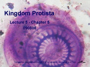
Kingdom Protista PowerPoint PPT Presentation
1 / 40
Title: Kingdom Protista
1
(No Transcript)
2
Kingdom Protista
- Includes Protozoa (primarily single-celled,
heterotrophic, eukaryotic organisms) AND some
autotrophic groups.
Kingdom Protista
Autotrophic Protista
3
Ecto-skeleton
Photosynthetic(autotrophic)
Colonial
Parasitic
Predatory
4
Kingdom Protista
- The Protozoa
- Phylum Kinetoplastida
- Phylum Ciliophora
- Phylum Apicomplexa
- Phylum Rhizopoda
For Wednesday 9/10
5
I. The protozoa
- Most are unicellular (some exceptions, i.e.
colonial or multicellular) - Protoplasmic grade of complexity all functions
take place in single cell or in each cell (if
colonial) - Most are microscopic (2 200 µm some
exceptions, i.e. Foraminiferans) - Marine, Freshwater, Terrestrial and Symbiotic
species
6
I. The protozoa
- The different groups of Protozoa can be
classified primarily by their modes of nutrition
and locomotion. - Mastigophora locomotion w/ flagella
- Ciliophora locomotion w/ cilia
- Sporozoa parasites w/ no obvious locomotory
structures - Sarcodina locomotion w/ pseudopodia
7
I. The protozoa
- Mastigophora - locomotion w/ flagella gt46,000
species - Ph. Euglenida
- Ph. Kinetoplastida
- Ph. Dinoflagellata
- Ph. Granuloreticulosa
- Ph. Diplomonadida
- Ph. Parabasilida
- Ph. Cryptomonada
- Ph. Choanoflagellata
- Ciliophora
- Ph. Ciliophora
- Sporozoa
- Ph. Apicomplexa
- Sarcodina
- Ph. Rhizopoda
8
Kingdom Protista
- The Protozoa
- Phylum Kinetoplastida
- Phylum Ciliophora
- Phylum Apicomplexa
- Phylum Rhizopoda
9
II. Phylum Kinetoplastida
- Characteristics
- Bodonids vs. Trypanosomes
- Agents of disease Leishmania, Trypanosoma
10
II. Phylum Kinetoplastida
- Characteristics
- Shape of cell difined by Pellicle (single layer
of microtubules beneath plasma membrane) - Flagella for locomotion (Bodonids w/ 2,
Trypanosomes w/ 1) - All possess a Kinetoplast (a disc-shaped, DNA
containing organelle within the mitochondrion)
Kinetoplast
Kinetoplast
11
II. Phylum Kinetoplastida
- Characteristics (cont.)
- Single nucleus
- Asexual reproduction by binary fission
- Complex Lifecycle monoxenous (1 host) or
heteroxenous (gt1 host)
A
A
(A) amastigote, (B) choanomastigote, (C)
promastigote, (D)
opisthomastigote,(E) trypomastigote.
location of the Kinetoplast (2) in relation to
the Nucleus (1) is key to differentiating among
lifecycle stages
A
A
P
A
P
P
P
P
P posterior endA anterior end
12
II. Phylum Kinetoplastida
- Characteristics
- Bodonids vs. Trypanosomes - The 600 described
species of Kinetoplastids can be divided into two
major subgroups. - Bodonids primarily free-living ? marine
freshwater habitats - Trypanosomes all parasitic ?invertebrates,
plants vertebrates.
Fig 5.11 Bodonid Trypanosome
13
II. Phylum Kinetoplastida
- Characteristics
- Bodonids vs. Trypanosomes - The 600 described
species of Kinetoplastids can be divided into two
major subgroups. - Bodonids primarily free-living ? marine
freshwater habitats - Trypanosomes all parasitic ?invertebrates,
plants vertebrates.
Trypanosomes are serious agents of disease in
humans and domestic animals
14
II. Phylum Kinetoplastida
- Characteristics
- Bodonids vs. Trypanosomes
- Agents of disease Leishmania, Trypanosoma
- Leishmania spp.
- All parasitic
- 5 species infect humans (including Leishmania
tropica, L. major, L. braziliensis, L. mexicana,
L. donovani) - Causes the disease leishmaniasis
- gt1 million humans infected annually kills 1000
people annually. - Heteroxenous lifecycle (transmitted by sandfly
vector)
15
Leishmania donovani - LIFECYCLE
In Sandfly Vector
In Vertebrate (mammal) host tissue
- Sandflies suck blood of infected animal ingest
amastigotes. - Amastigotes develop in gut of sand fly develop
into promastigotes. - Promastigotes migrate to pharynx.
- Sandfly injects promastigotes into vertebrate
while taking blood meal.
- Parasite lives in macrophages of vertebrate
blood, spleen, liver.
All Figures from Roberts Janovy, Foundations of
Parasitology, 7th edition 2005
16
Cutaneous Leishmaniasis
L. mexicana lesions chronic
L. major lesion bleeds quickly and is of short
duration.
L. tropica lesion is dry and persists for months.
In both species, the lesions eventually dry up to
produce a depressed, unpigmented scar.
Heals well, except when lesions on poorly
vascularized ear
Mucocutaneous Leishmaniasis
Visceral Leishmaniasis
L. braziliensis lesion involves nasal system,
causing degeneration of cartilaginous soft
tissues.
L. donovani causes great enlargement of spleen
and liver
Leads to great upper lip, palate and gum
deformity.
Death rarely spontaneous recovery.
17
II. Phylum Kinetoplastida
- Characteristics
- Bodonids vs. Trypanosomes
- Agents of disease Leishmania, Trypanosoma
- Trypanosoma spp.
- All parasitic, occur in all classes of
vertebrates. - T. brucei causes non-lethal nagana in African
hoofed animals fatal to livestock. (impossible
to raise livestock on 45 million miles2 of
Africa) - T. brucei gambiense and T. brucei rhodesiense
cause sleeping sickness in humans (Kills 65,000
people annually). - T. cruzi causes Chagas disease in humans
(Infects 15-20 million people at any given time).
18
Trypanosoma brucei gambiense - LIFECYCLE
15-35 days
- Glossina (Tsetse fly vector) suck up parasites
in when taking a blood meal from an infected
vertebrate host. - The parasites locate in the
posterior section of the insects midgut.
19
Trypanosoma brucei gambiense - LIFECYCLE
- Trypomastigotes migrate to the foregut, then
pharynx where trypomastigotes transform into
choanomastigotes. - The choanomastigotes
undergo asexual reproduction in the salivary
glands. - Finally transforming back into
metacyclic trypomastigotes.
20
Trypanosoma brucei gambiense - LIFECYCLE
- Finally, the infective trypomastigotes are
injected into the vertebrate host while the
tsetse takes a blood meal. - Once in the
vertebrate host, the parasites multiply as
trypomastigotes in the blood and lymph.
21
Trypanosoma brucei gambiense -
- Symptoms of African Sleeping sickness
- Small sore at site of inoculation
- Swollen lymph nodes (groin, neck, legs)
- Invasion of central nervous system causes mental
dullness, increased sleepiness, coma and death.
22
Trypanosoma cruzi - LIFECYCLE 10 days
When reduviid bugs feed they often defecate on
the skin of their host. Feces may contain
metacyclic trypanosomes, which gain entry into
the body of a vertebrate host through a bite,
scratched skin, or mucous membranes that are
rubbed with fingers contaminated with insects
feces. (Roberts Janovy, 2006)
23
Trypanosoma cruzi - LIFECYCLE 10 days
Amastigotes are likely to form pseudocysts in
tissues such as heart muscle (and sometimes
brain). Trypomastigotes will be in the host
blood.
24
Trypanosoma cruzi -
Romanas sign
- Symptoms of Romanas sign
- swelling (edema) of eye if bug feces are rubbed
into eye - Symptoms of Chagas disease
- muscle tone in organs (heart, colon, esophagus)
is destroyed, as nerve ganglia are destroyed by
pseudocyst rupture and inflammation.
Megacolon
25
Kingdom Protista
- The Protozoa
- Phylum Kinetoplastida
- Phylum Ciliophora
- Phylum Apicomplexa
- Phylum Rhizopoda
26
III. Phylum Ciliophora
- 12,000 described species
- Benthic (bottom) planktonic (float, drift,
swim) - marine, brackish, freshwater damp soils
- free-living, ectosymbionts, endosymbionts,
parasitic
27
III. Phylum Ciliophora
- Characteristics
28
III. Phylum Ciliophora
- Characteristics
- Shape of cell maintained by pellicle fibrillar
structures. - Cilia for locomotion (somatic ciliature)
feeding (oral ciliature). single simple cilia
grouped compound cilia (e.g., cirri)
Fig 5.14
Fig 5.13
- Cilia are in rows (kineties)
- Each cilium is anchored by three fibrillar
structures ( postcilliary microtubules
transverse microtubules a
kinetodesmal fiber)
29
Locomotion / Somatic Ciliature
30
Locomotion / Somatic Ciliature
Fig 5.13 Paramecium 5.15 Helical pattern of
forward movement of Paramecium.
Video clip?
31
Locomotion / Somatic Ciliature
Fig 5.15 Hypotrich ciliates cirri are used to
crawl or walk over surfaces
Fig 5.13 Hypotrich ciliate with cirri
32
Feeding / Oral Ciliature
Cytopharynx everts, sticks to prey, then inverts
back into cell, thus pulling the prey into the
food vacuole.
Fig 5.17 Holozoic feeding in ciliates. Didinium
consuming Paramecium.
Fig 5.18 Feeding currents produced by two
ciliates. (A) Euplotes (B) Stentor. The ciliary
currents bring suspended food to the cytostome (
cell mouth) where it can be ingested.
33
See Video clip of stalked ciliates feeding
Parasitic crustaceans, from Nebraska minnows,
covered with commensal ciliates.
34
Feeding / Oral Ciliature
Fig 5.20 Suctotorians lack cilia as adults. (A)
They use extrusomes called Haptocysts which are
discharged from tips of tentacles when come into
contact with prey. (B) Following attachment to
prey, a temporary tube forms within the tentacle
and (C) contents of prey are sucked into the
tentacle and (D) incorporated into food vacuoles.
35
Feeding / Oral Ciliature
Bolek
Endosymbiotic ciliates break down grasses eaten
by host. Other cattle rumen ciliates feed on
bacteria some prey on other ciliates. These
are the cattle rumen ciliate Entodinium
caudatum.
Bolek
Note kineties surface architecture
Bolek
36
III. Phylum Ciliophora
- Characteristics (cont.)
- 2 nuclei types macronucleus micronucleus.
- Asexual and sexual reproduction.
- Macronucleus
- controls general functions of the cell
- many sets of chromosomes ( hyperpolyploid)
- Micronucleus
- reservoir of genetic material in cell
- two sets of chromosomes (diploid)
37
Asexual reproduction
- Transverse binary fission in Paramecium
- careful mitotic division of micronucleus to
ensure equal distribution of daughter
micronuclei to progeny of the division. - structures not centrally or symmetrically placed
in cell must be generated after fission. - Colonial and solitary ciliates
- Vorticella divides, both remain attached to
colony - Ephelota, a solitary ciliate, young buds are
ciliated and swims off.
38
Sexual reproduction / Conjugation
Each original conjugant results is 4 new diploid
daughter organisms.
to haploid condition
still haploid
now micronucleus diploid (synkaryon)
still haploid
39
Take-home Messages - 090808
- The different groups of Protozoa can be
classified primarily by their modes of nutrition
and locomotion. - Parasitic Kinetoplastids are serious agents of
disease in humans, particularly Leishmania spp.
Trypanosoma spp. - Ciliates are exceedingly diverse in their
habitats, locomotion, feeding strategies, and
reproductive mode.
40
Study Questions - 090808
- What are the four major groups within the
protozoan classification system? Give the
defining character for each. - Describe the lifecycle of Trypanosoma brucei
gambiense. - What portion of the lifecycle, in the organism
that causes Chagas disease, is responsible for
the tissue damage of the heart, colon, and
esophagus? - Describe feeding in Suctotorians.
- What are the fates of the micronucleus and
macronucleus following transverse binary fission
in Paramecium?

