Studying the phagocytosis of apoptotic cells - PowerPoint PPT Presentation
1 / 41
Title:
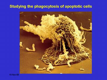
Studying the phagocytosis of apoptotic cells
Description:
Actin-dependent process allowing a cell to engulf and digest large particulate ... e.g. lamellocytes and encapsulation of parasitic wasp eggs. ... – PowerPoint PPT presentation
Number of Views:309
Avg rating:3.0/5.0
Title: Studying the phagocytosis of apoptotic cells
1
Studying the phagocytosis of apoptotic cells
2
Phagocytosis
Actin-dependent process allowing a cell to
engulf and digest large particulate matter (gt
1um) Very important for host defence
Differs from Endocytosis clathrin-dependent
uptake of macromolecules and small
particles. Pinocytosis uptake of solutes into
the cell can involve clathrin.
3
Related processes
4
Endocytosis e.m. analysis
Phagocytosis requires similar membrane
internalisation events BUT does not
necessarily involve clathrin
5
Phagocytosis
First described by Metchnikoff in 1880s
microscopic observation of starfish larvae and
also in higher organisms.
Process is largely conserved at the molecular
level from primitive organsisms (e.g.
Dictyostelium) to vertebrates. In Drosophila
there are specialised phagocytic cells
(hemocytes) these cells are important in host
defence e.g. lamellocytes and encapsulation of
parasitic wasp eggs. Phagocytosis occurs in
Caenorhabditis important for clearance of dying
cells during worm development.
6
Many cells capable of phagocytosis in
vertebrates Non-professional phagocytes Fibrobl
asts, epithelial cells, B cells. Professional
phagocytes Monocytes/macrophages Kupffer
cells Osteoclasts Alveolar macrophages Microglial
cells Neutrophils Immature dendritic cells.
phagocytosis of Candida
Important for clearance of pathogens, apoptotic
cells and cellular debris. Phagocyte responses
can influence progression of inflammation, tissue
remodelling and development of immune responses.
7
Steps involved in phagocytosis
Recognition and adhesion Formation of phagocytic
cup Membrane extension around particle Fusion
of phagosome with lysosomes Particle degradation
8
Steps involved in phagocytosis
9
Phagocytosis target recognition
Membrane receptors involved non-opsonin Pattern
recognition receptors TLRs Lectin-like receptors
e.g. DEC205 Mannose receptors Scavenger
receptors Some integrins e.g. aMb2 binds
Neisseria gonnerheae aVb3 binds Bordetella
and Adenovirus opsonin Antibodies Complement
components direct opsonins and complement
activation proteins LPS binding proteins
10
Opsonic receptors Antibodies
- Antibody recognised by several classes of FcR
receptor binding can trigger internalisation. - FcgR 3 receptors
- all Ig superfamily
- CD16 (FcRIII), CD32 (FcRIIa and b) both low
affinity recognise multimeric IgG - CD64 (FcRI) high affinity recognises monomeric
IgG too. - Ligand binding can trigger respiratory burst e.g.
FcRIIa via ITAMs - Or can inhibit responses e.g. FcRIIb via ITIMs
- FcaR 3 isoforms
- CD89 - again Ig superfamily
- Again ligand binding can promote respiratory
burst activity. - FceR low affinity (as opposed to high affinity
on basophils/mast cells - CD23 has C-type lectin domain.
- Ligand binding can trigger inflammatory mediator
release
11
Opsonic receptors Complement
- Phagocytosis generally requires cellular
activation - Complement recognised by several different types
of receptor - CR1 (CD35) recognises C3b, C3dg
- -Mediates adhesion of target to phagocyte
- aMb2 recognises iC3b
- aXb2 recognises iC3b
- Ligand binding can trigger internalisation
signals for other CR.
12
FcR v CR mediated phagocytosis
13
How is phagocytosis controlled?
Clustering of FcR Phosphorylation of ITAM motifs
by Src kinases Recruitment of Syk kinase Syk
phosphorylates PI3K 85kDa Generates IP3 near to
clustered receptors Also recruitment of SH2
containing proteins e.g. SHP1, Gab3, SLP-76 etc.
14
How is phagocytosis controlled?
15
How is phagocytosis controlled?
Differential localisation of signalling molecules
during internalisation process.
16
EE early endosome LE - late endosome Ly -
lysosome
Desjardins NRI 3 280
17
Therapeutic Strategies for inflammatory
diseaseinduction of apoptosis
Neutrophil apoptosis
Phagocytic Motile granules contain cationic
proteins oxidants enzymes inflammatory mediators
Effector function uncoupled Membrane integrity
retained Granule contents intact
Inflammation e.g. pneumonia massive inflammatory
cell recruitment
18
Regulation of apoptosis
INHIBITION
PROMOTION
TNF-a Fas ligation NO Phagocytosis Bacterial and
fungal products
LPS, C5a, GM-CSF, IL-1b, IFNg, LT B4, hypoxia,
glucocorticoids, Ca 2 i, cAMPi
Cleared by macrophages.
19
Triggered apoptosis
20
Potential for therapy?
- Selectively drive granulocyte death
- With Glucocorticoids
- With TNF-alpha or Fas ligation together with NFkB
block - rapid acceleration of granulocyte death
- However...
- Failure to clear increased numbers of apoptotic
cells may compromise the resolution of
inflammation
21
Therapeutic Strategies for inflammatory
diseaseRegulation of cell clearance
Inflammation Pneumonia
functional down-regulation contents retained
phagocytosis
22
Video of monocyte-macrophage phagocytosis
23
Apoptotic cell clearance anti-inflammatory
Altered cell surface molecules
Opsonins
Modified or apoptotic lipid/CHO/ICAM-3
Ox-LDL
PS
TSP-R?
iC3b
C1q
?
CD51/61 CD36 TSP
C1qR
PS receptor
CD36
LOX-1
CD14
CD11b CD18
CD29
lectin
scavenger receptor
24
Many receptors involved in macrophage recognition
of apoptotic cells
Receptors involved PSR? Scavenger receptors
CD36, SRA CD91 CD14
Altered cell surface molecules Phosphatidylserine
Carbohydrate AGE? Lipids oxidised or
modified lipids? Proteins - ICAM-3?
Opsonins C3b - C1q - TSP - ? PS binding -
MFG-E8 Gas6
Receptors CD11b/CD18 C1qR CD51/CD61 CD36 Mer
25
Genes identified as being important for
phagocytosis of cellular corpses in C. elegans
Images from Zheng Zhou Baylor College
26
Many of the proteins implicated in phagocytosis
also important for adhesion and migration
27
Macrophage phagocytosis of apoptotic neutrophils
Apoptotic neutrophils present in trypsinised
macrophages and are internalised in e.m. analysis
Phagocytosis can be quantified by microscopy or
by flow cytometry
28
Macrophage populations for study
- Monocyte-derived macrophages (human)
- Monocyte isolated and cultured in vitro to
acquire macrophage characteristics - Alveolar Macrophages (human/animal)
- From bronchoalveolar lavage
- Peritoneal Macrophages (animal)
- Either resident or elicited with inflammatory
agent - Bone-marrow-derived macrophages (animal)
- Expanded from progenitors over 7-10 days
29
How can we measure phagocytosis?
- Microscopy
- Tedious
- Time consuming
- Observer bias
- Difficult to be certain of particle
internalisation
30
How can we measure phagocytosis?
- Flow cytometry
- Rapid
- Cell by cell analysis
- Observer bias eliminated
- Still can be difficult to distinguish
internalisation from binding
31
Labelling neutrophils does not affect
characteristics of apoptosis
B
A
unlabelled
CMFDA
ns
Percent CD16-Hi
ns
Percent CD62L
Fluorescence
ns
Percent Annexin V
Apoptotic Neutrophils
Neutrophils
32
Macrophages can be identified by laser scatter
properties
Side Scatter
R1
LC
Forward Scatter
33
Gates for sorting cell populations
A
R1
Fluorescence
R2
Forward Scatter
34
Sorted populations
35
Flow cytometry confirms that apoptotic cells are
internalised
CMFDA neutrophils control PECy5
Macrophages CD14 PECy5
CMFDA neutrophils CD15 PECy5
Macrophages that have phagocytosed apoptotic
neutrophils CD15 PECy5
CMFDA fluorescence
Macrophages CD15 PECy5
PECy5 fluorescence
36
Analysis of known factors that influence
phagocytosis
60
Percent FL1-positive macrophages
40
20
0
Baseline
CD44
EDTA
Cytochalasin D
4
0
C
Treatments
37
The effects of washing cell loss and percentage
phagocytosis
A
phagocytosis
No wash
1 Wash
2 Washes
3 Washes
B
phagocytosis
No wash
1 Wash
2 Washes
3 Washes
Number of Washes
38
Assay applicable to other cell types
B
R1
Macrophages
A
CMFDA-labelled
Side scatter
unlabelled
Contaminating
LC
Viable
Mutu I Cells
Apoptotic
Mutu I Cells
C
R2
Fluorescence
Forward scatter
39
Triggering CD44 can influence clearance in vivo
- Mice injected with CD44 mAb
- 15 minutes later 20x106 CFDA labelled neutrophils
injected - after 7 minutes peritoneal cavity lavaged
- F4/80 positive macrophages that have ingested
apoptotic cells determined
83 7
70 25
Untreated CD44 treated
Potential for rescue of phagocytic defects?
40
Phagocyte responses to apoptotic cells
Down-regulation of pro-inflammatory cytokine
(e.g.TNFa release in response to LPS and other
stimuli Release of TGF-b Release of IL-10?
41
Finally - cross-presentation exogenous antigen
into class I


