A Pyro-Electric Crystal Particle Accelerator - PowerPoint PPT Presentation
1 / 1
Title:
A Pyro-Electric Crystal Particle Accelerator
Description:
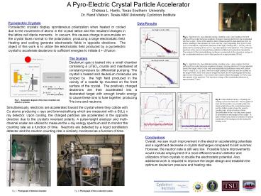
Fig. 2. Photograph of theInner Chamber. Fig. 3 Photograph of the ... Discontinuities in the x-ray rate were caused by spontaneous discharges of the crystal. ... – PowerPoint PPT presentation
Number of Views:49
Avg rating:3.0/5.0
Title: A Pyro-Electric Crystal Particle Accelerator
1
A Pyro-Electric Crystal Particle
Accelerator Chelsea L. Harris, Texas Southern
University Dr. Rand Watson, Texas AM University
Cyclotron Institute
Pyroelectric Crystals Pyroelectric crystals
display spontaneous polarization when heated or
cooled due to the movement of atoms in the
crystal lattice and the resultant changes in the
lattice cell dipole moments. In vacuum, this
causes charge to accumulate on the crystal faces
normal to the polarization, producing a large
electrostatic field. Heating and cooling
generate electrostatic fields in opposite
directions. The object of this work is to
utilize the electrostatic field produced by a
pyroelectric crystal to accelerate deuterons to
sufficient energies to initiate d d fusion.
Data/Results
Fig. 4. Spectrum of x rays detected during a
heating cycle. Upon heating, the front surface of
the crystal becomes positively charged, causing
electrons to be accelerated into the Cu disc.
Some of the electron-atom collisions eject
K-shell electrons from Cu atoms, resulting in the
emission of Cu K? and K? x rays originating from
2p to 1s and 3p to 1s transitions, respectively.
Because of the high counting rate (? 20 K/s),
pile-up peaks due to summing of two Cu K x rays
also appear in the spectrum. The continuous
spectrum is the due to bremsstrahlung radiation
emitted as the electrons decelerate. The endpoint
of the bremsstrahlung continuum corresponds to
the maximum energy of the accelerated electrons,
and in this spectrum it is approximately 88 keV.
The System Deuterium gas is leaked into a small
chamber containing a LiTaO3 crystal and
maintained at constant pressure by differential
pumping. The crystal Is heated and deuterium
molecules are ionized by the high field produced
in the vicinity of a needle tip mounted on the
front surface of the crystal. The positively
charged deuterons are then accelerated into a
deuterated target with enough kinetic energy to
cause these ions to fuse together, producing 3He
ions and neutrons.
Fig. 5. Spectrum of x rays detected during a
cooling cycle. Upon cooling, the front surface
of the crystal becomes negatively charged,
causing electrons to be accelerated toward the
target, which in this case was deuterated
polyethylene mixed with carbon. The K x rays of
Zn, Cd, and S are from a light coating of cadmium
and zinc sulfide on the target wheel, which was
used to image the beam (as in the photograph at
the top right corner of the poster). Iron screws
were used to attach the targets, so Fe Kx rays
also appear in the spectrum. The maximum energy
of the accelerated electrons is approximately 70
keV.
D D ? 3He n (820 KeV)
(2.45 MeV) D D ? T p
(1.01 MeV) (3.02 MeV)
Fig. 6. Data obtained during a heating and
cooling cycle in two test runs. The top graph of
each set shows the x-ray counting rate, the
center graph shows the crystal temperature, and
the bottom graph shows the neutron counting rate,
all as a function of time. Discontinuities in
the x-ray rate were caused by spontaneous
discharges of the crystal. In both of these
runs, there is evidence of neutron bursts above
the background rate during the heating cycle.
Fig. 1. Schematic diagram of the inner chamber
and detector systems.
Simultaneously, electrons are accelerated toward
the crystal where they collide with Cu atoms
producing x rays and bremsstrahlung which are
measured with a Si(Li) x ray detector. Upon
cooling, the charged particles are accelerated in
the opposite direction due to the crystals
reversed polarity. A pulse-height analyzer and
multi-channel scalar are utilized to measure the
x-ray energy spectrum and to monitor the counting
rate as a function of time. Neutrons are
detected by a liquid scintillation detector and
the neutron counting rate is similarly monitored
as a function of time.
Conclusions Overall, we saw much improvement in
the electron accelerating potentials and a
significant decrease in crystal discharges
compared to last summer. However, the neutron
rate is still very low. Possible future
improvements would include employment of a more
efficient neutron detector and utilization of two
crystals to double the electrostatic potential.
Also, additional work is required to improve the
target design and establish the optimum deuterium
pressure and heating rate.
Fig. 2. Photograph of theInner Chamber
Fig. 3 Photograph of the accelerator system.


