Technical BioImaging PowerPoint PPT Presentation
1 / 7
Title: Technical BioImaging
1
Technical BioImaging
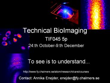
TIF045 5p 24th October-9th December
- To see is to understand...
http//www.fy.chalmers.se/atom/research/cars/cours
es
Contact Annika Enejder, enejder_at_fy.chalmers.se
2
Technical BioImaging
Aims
- To understand how a 3-dimensionell, highly
resolved image is created in a modern light
microscope - The physics behind light-matter interaction in a
microscopic probe volume - How cell structures and even biomolecules can be
imaged and manipulated in a microscope - Give a flavor of all the novel microscopy
techniques currently emerging within the exciting
inter-disciplinary field of physics and molecular
cell biology
Contact Annika Enejder, enejder_at_fy.chalmers.se
3
Technical BioImaging High-lights from the
program
- The Nikon Microscopy Workshop at Chalmers
University - 26-27th October
- From the fundamentals on the anatomy of a modern
microscope to hands-on training in how to use a
microscope collect images of biological samples
Contact Annika Enejder, enejder_at_fy.chalmers.se
4
Project conducted on the research microscopes at
The Centre for BioPhysical Imaging14-18th
November
Technical BioImaging High-lights from the
program
- Confocal microscopy of GFP labeled proteins
- 2-photon fluorescence microscopy
- Fluorescence Lifetime Imaging FLIM
- CARS - Coherent Anti-Stokes Raman Scattering
microscopy - Vibrational (Raman) microscopy
- Surface Plasmon Resonance and Nanoparticle
sensors - Optical tweezers and manipulation
5
Technical BioImaging High-lights from the
program
- Lecture series by Prof James B Pawley
- University of Wisconsin, Madison, USA
- 21-23th November
The editor of the course literature Handbook of
Biological Confocal Microscopy
6
Technical BioImaging Course Contents
- The light microscope has traditionally been our
most important instrument to catch a glimpse of
the fascinating micro-cosmos of biological cells.
The fundamental optics of the microscope and its
function to image with an ever increasing
magnification and resolution has been
well-known for several hundred years. However,
the information provided by the conventional
snapshots of transmitted light is no longer
enough within the modern biosciences in order to
gain full understanding of the complex processes
life comprises. The requirements on resolution
and contrast in order to visualize the
invisible have the past decade drastically been
changed we now want to be able to monitor
biochemical processes in living cells on
molecular level and with high temporal resolution
rather than merely visualizing cellular
structures. This has motivated physicists in
close collaboration with biologists to develop a
range of sophisticated microscopy methods, where
the conventional light source is replaced by
technically advanced laser systems.
Micro-spectroscopic analysis and imaging of
biomolecules and molecular markers in the sample
is done by inducing fluorescence and vibrational
processes. Living cells are captured in the probe
volume and manipulated with laser tweezers. The
light emitted from the sample, sometimes only
single photons, is wavelength-selectively
filtered and projected onto a sensitive detector.
An image is formed and finally analyzed by means
of powerful image analysis software. These
advanced laser-based imaging systems set high and
new requirements on the user of modern
microscopes, one of which is good knowledge in
physics. Here we intend to bring up concepts from
a wide range of fundamental physics courses and
use them to understand how a 3-dimensionell
picture is created in a modern light microscope,
how cell structures and biomolecules selectively
can be imaged and manipulated in fluorescence and
spectroscopically based microscopes, and how
important image analysis and sample preparation
is for the final results.
Contact Annika Enejder, enejder_at_fy.chalmers.se
7
Technical BioImaging
TIF045 5p
For further information, contact Annika Enejder
enejder_at_fy.chalmers.se
http//www.fy.chalmers.se/atom/research/cars/cours
es

