Hearing PowerPoint PPT Presentation
1 / 13
Title: Hearing
1
Hearing
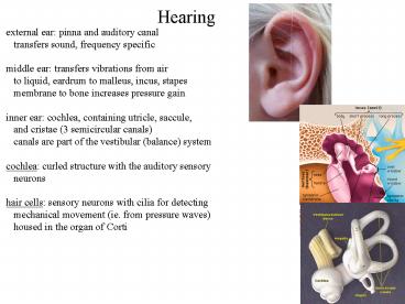
external ear pinna and auditory canal
transfers sound, frequency specific middle ear
transfers vibrations from air to liquid,
eardrum to malleus, incus, stapes membrane to
bone increases pressure gain inner ear cochlea,
containing utricle, saccule, and cristae (3
semicircular canals) canals are part of the
vestibular (balance) system cochlea curled
structure with the auditory sensory
neurons hair cells sensory neurons with cilia
for detecting mechanical movement (ie. from
pressure waves) housed in the organ of Corti
2
Hearing
hair cells have a receptor (microvilli) apical
side of various lengths and basal
(connection) side for synapse formation microvill
i are linked together, and move in unison,
regulating a nonselective cation
channel displacement toward long hairs open
channels/depolarizing displacement toward short
hairs closed channels/hyperpolarizing depolariza
tion more synaptic release hyperpolarization
less synaptic release fastest gating of any
sensory system required for high frequency
sounds
3
Hearing
low frequencies are detected at the basal end of
organ of Corti high frequencies are detected at
the apical end of organ of Corti outer hair
cells actually change their shape using voltage
gated motors has a unique membrane bound motor
protein family (Prestin) shape changes the
electrical conductivity of the OHC capacitance
changes of the membrane motor/shape filter
frequency changes and increases the response
of the neuron motors have a voltage sensing
helix and are all on lateral side of cell
4
Hearing
different regions of the cochlea have different
extracellular fluids giving rise to different
currents in different parts of the hair
cell perilymph normal extracellular medium near
hairy end of cell endolymph high potassium
extracellular fluid around rest of the hair cell
increases the sensitivity of the hair cell
using K channels reduces blood flow and
energy requirements, reducing 'noise' intense
sound can kill hair cells, as can some
antibiotics some loss can be restored using
cochlear implants, a microphone with
electrical implants along the cochlea
5
Hearing
spiral ganglion (2 neuron types) send bipolar
axons to cochlea and CNS type I connect to a
single hair cell and are myelinated, and very
fast type II connect to many hair cells, are not
myelinated, and are slow both go to the cochlear
nucleus of the brainstem neurons are frequency
selective narrow tuning in sensitive regions
broader tuning in insensitive ones tonotopic
mapping lowest frequency fibers innervate
apical positions and high frequency basal ones
6
Hearing
louder sounds give broader response ie. nearby
frequency neurons fire sensitivity of
different fibers varies-- range of responses up
to 70 dB 3 different patterns of spontaneous
activity are found in auditory fibers low rates
(lt0.5/second) more terminals, low
sensitivity medium (gt0.5 to 17.5/second) many
terminals, moderate sensitivity high rates
(gt17.5/second) fewer terminals, highest
sensitivity
7
Hearing
auditory masking blocking out one signal using a
different signal occurs by several
mechanisms adaptation high initial rates of
firing, then lowering frequency over time
occurs at the hair cell or auditory nerve level
blocks later stimuli because the firing
frequency is already reduced suppressive masking
or 2-tone masking, uses 1 frequency to block the
response at another frequency generated
because of the membrane covering the organ of
Corti
8
Hearing
superior olivary complex sends out ennervation to
the cochlea 2 types medial ennervating outer
hair cells, lateral synapse on type I medial
fibers use acetylcholine to hyperpolarize hair
cells, reducing sensitivity to soft, low
intensity sounds and reducing loud
sounds superior olivary complex is responsible
for positional sound information
9
CNS Auditory Features
auditory processing in the CNS are generally
tonotopically organized later processing of
the frequency maps gives desired properties bats
or other echolocating species have expanded
sensitivity in that range of
hearing tonotopic maps maintain plasticity to
deal with hearing loss- important for age
related hearing loss loss starts at high
frequency, middle frequencies then expand
10
CNS Auditory Features
cochlear nucleus has numerous types of neurons
based on firing patterns in single cells, each
with particular morphologies as well different
firing patterns corellate with different
functional processing steps and initiates
parallel processing of auditory
information various patterns are generated by
different inhibitory or excitatory
interneurons other neurons may be monaural or
biaural- integrates positional information by
time delay brainstem is generally excited by
the contralateral side- crosses midline
11
CNS Auditory Features
inputs from brainstem regions to higher regions
go through the inferior colliculus, which
maintains tonotopic mapping but receives
different input in different layers/nuclei of
the inferior colliculus inferior colliculus maps
onto the superior colliculus, integrating visual
perception space maps and tonotopic position
maps inferior colliculus connects tonotopic
cortex through the medial geniculate nucleus
4 regions of the cortex have tonotopic maps,
but others do not non-tonotopic cortex gets
input from the dorsal geniculate geniculate-am
ygdala pathway mediates fear conditioning
12
CNS Auditory Features
cortex- highest processing level lateral
geniculate- cortex switching station inferior
colliculus- links to visual system in superior
colliculus/tectum cochlear nucleus- initial
CNS spiral ganglion target ear/hair cells and
spiral ganglion
13
CNS Auditory Features
cortical columns are vertical orientations of
processing through all 6 layers of the cortex
where layer neurons in column have similar
patterns columns generally respond to either
binaural or tonotopic, but not both cells
within columns have large projections to
contralateral cortex, allowing 1 ear to
control processing, often found with A1 space
maps several regions of cortex respond to
complex species-specific sounds in primates,
left cortex lesions cause larger defects than
right cortex language is particularly linked to
2 regions, Wernicke's and Broca's simple
sounds are processed in auditory cortex complex
ones in association areas of the cortex

