Objective PowerPoint PPT Presentation
1 / 1
Title: Objective
1
The use of Computed Radiography for Megavoltage
Linear Accelerator Quality Control I Patel, T
Natarajan and MC Kirby North Western Medical
Physics, Radiotherapy Department, Rosemere Cancer
Centre, Royal Preston Hospital, Preston, UK
Objective One of the major developments in UK
hospitals is a national programme for integrating
IT systems. Part of this, is the full roll-out
of PACS/digital imaging to all radiology and
radiotherapy departments which involved moving
simulator and film-based portal imaging onto a
Computed Radiography (CR) platform. The Kodak
2000RT CR system (figure 1) was chosen, since it
gives excellent clinical images, but also has an
established package for IMRT type dosimetry with
small (24 x 30cm) plates1. We wanted to gain
maximum benefit from the system by examining how
it could be used as a simple film substitute for
performing standard linear accelerator quality
control (QC). However many standard QC fields
need a large (gt 30 x 40 cm) imaging area2.
EPIDs, with their smaller active field of view,
may be used, but the fields must be subdivided.
This is impractical for CR, where plates still
require processing before the image is
available digitally. This paper describes our
experiences with commissioning and implementing
large (35 x 43 cm) CR plates for this purpose.
Methods and Materials We examined the
properties of three (35x43 cm) Agfa CR imaging
plates (model MD10) processed with the Kodak
Radiation Oncology Beam Dosimetry package (v1.0)
for a variety of QC procedures on our six Elekta
(Elekta Ltd, Crawley, UK) linacs. The QC
procedures included fields (figure 2) for
examining MLC alignment, centring and
magnification, X-ray/light field congruence for
both leaves and backup collimators, multisegment
fields for MLC leaf and backup collimator
calibration (for both standard 1 cm and 0.4 cm
MLCs). Experiments were conducted as
simultaneous dmax, isocentric irradiation of CR
plates and film (X-omat-V and EDR2, Kodak,
Rochester, NY), for 6MV X-rays. Exposures were
made to examine dose density response,
uniformity, reproducibility of scaling and signal
decay with time. Two scanning resolutions were
investigated - 1024 and 2048 pixels, although
most scans were performed at the lower
resolution. RIT 113 (v4.4) (RIT Inc., Colorado
Springs, CO) software was used for analysis using
principally its ROI and line profiles
functionality (figure 3). Films were analysed
using current methods of manual densitometer or
RIT software (for the Bayouth test only).
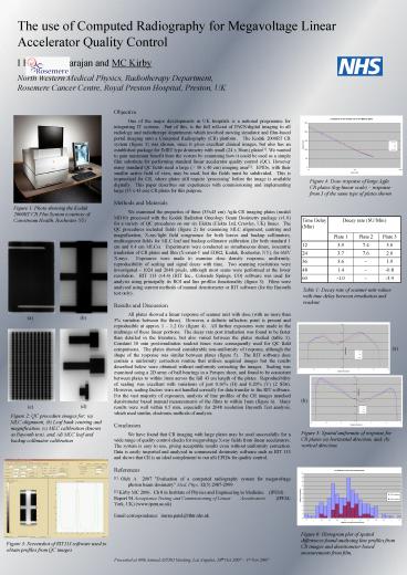
Results and Discussion All plates showed a
linear response of scanner unit with dose (with
no more than 5 variation between the three).
However, a definite inflection point is present
and reproducible at approx 1 1.2 Gy (figure 4).
All further exposures were made in the midrange
of these linear portions. The decay rate post
irradiation was found to be faster than detailed
in the literature, but also varied between the
plates studied (table 1). Constant 10 min
post-irradiation readout times were consequently
used for QC field comparisons. The plates showed
considerable non-uniformity of response, although
the shape of the response was similar between
plates (figure 5). The RIT software does contain
a uniformity correction routine that utilises
acquired images but the results described below
were obtained without uniformity correcting the
images. Scaling was examined using a 2D array of
ball bearings in a Perspex sheet, and found to be
consistent between plates to within 1mm across
the full 43 cm length of the plates.
Reproducibility of scaling was excellent with
variations of just 0.16 (H) and 0.20 (V) (2
SDs). However, scaling factors were not handled
correctly for data transfer to the RIT software.
For the vast majority of exposures, analysis of
line profiles of the CR images matched
densitometer based manual measurements of the
films to within 1mm (figure 6). Many results
were well within 0.5 mm, especially for 2048
resolution Bayouth Test analysis, which used
similar, electronic methods of analysis.
Conclusion We have found that CR imaging with
large plates may be used successfully for a wide
range of quality control checks for megavoltage
X-ray fields from linear accelerators. The
system is easy to use, giving acceptable results
even without uniformity correction. Data is
easily imported and analysed in commercial
dosimetry software such as RIT 113 and shows that
CR is an ideal complement to our aSi EPIDs for
quality control. References 1 Olch A 2007
Evaluation of a computed radiography system for
megavoltage photon beam dosimetry Med. Phys.
32(5) 2987-2999 2 Kirby MC 2006. Ch 8 in
Institute of Physics and Engineering in Medicine
(IPEM) Report 94 Acceptance Testing and
Commissioning of Linear Accelerators (IPEM,
York, UK) (www.ipem.ac.uk) Email correspondence
imran.patel_at_lthtr.nhs.uk
Table 1 Decay rate of scanner unit values with
time delay between irradiation and readout.
Presented at 49th Annual ASTRO Meeting, Los
Angeles, 28th Oct 2007 1st Nov 2007

