
Microscope PowerPoint PPT Presentation
1 / 2
Title: Microscope
1
Microscope
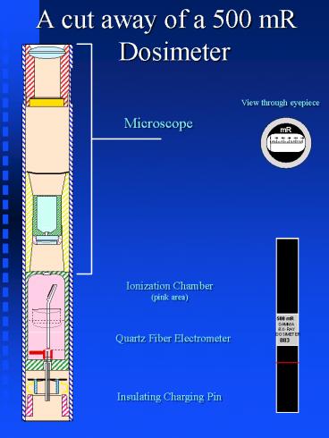
- A cut away of a 500 mR Dosimeter
View through eyepiece
Ionization Chamber (pink area)
500 mR GAMMA X-RAY DOSIMETER 883
Quartz Fiber Electrometer
Insulating Charging Pin
2
Pocket DosimetersHow do they work?
- They are zeroed with a dosimeter charger at the
Insulating Charging Pin. This device puts a
positive charge on the Quartz Fiber Electrometer.
This charge pushes the Quartz Fiber away from
the Electrometer. - As the Ionization Chamber is exposed to Gamma or
X-rays, negative ions are attracted to the
positively charged Electrometer. This attaction
bleeds off the Electrometer charge, causing the
Quartz Fiber to move toward the Electometer. - A meter scale is located after the front lens of
the Microscope. When viewed through the
eyepiece, the meter scale and Quartz Fiber appear
as one and indicate the exposure. - The meter indication will continue to increase
until the positive charge is removed from the
Electometer, at which time the dosimeter must be
rezeroed.
Recommended
