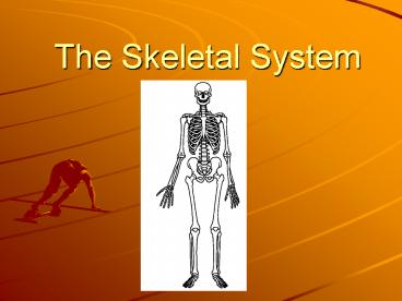
The Skeletal System PowerPoint PPT Presentation
1 / 46
Title: The Skeletal System
1
The Skeletal System
2
Functions
- Supports the body (Like the frame of a house)
- Protects internal organs (skull protects brain,
ribs protect the lungs) - Provides for movement (uses a lever system)
- Stores mineral reserves (calcium)
- Provides a site for blood cell formation
3
- How many bones does a human have?
4
- How many bones does a human have?
- 206
5
4 types of bones
- Long found in arms and legs
6
- Short wrists and ankles
7
- 3. Flat shoulder blades, cranium
8
- 4. Irregular vertebrae
9
Skeleton is divided into two parts
- Axial skeleton supports the central axis of the
body. Consists of the skull, the vertebral
column, and the rib cage. - Appendicular skeleton- bones of the arms and
legs, pelvis and shoulder area
10
(No Transcript)
11
Are bones living tissue?
- Yes!! Bones are a solid network of living cells
and protein fibers that are surrounded by
deposits of calcium salts.
12
Structure of bones
- Periosteum tough layer of connective tissue
that surrounds the bone - Contains blood vessels that carry oxygen and
nutrients to the bone - Growth, repair and development is started in this
membrane.
13
(No Transcript)
14
Two types of bone (osseous) tissue
15
- Compact bone layer beneath the periosteum.
Dense layer, but not solid. - Running through compact bone are the Haversian
canals which are a series of tubes that contain
blood vessels and nerves.
16
- Spongy bone found inside the outer layer of
compact bone. Less dense tissue. - Found in the ends of long bones and in the middle
of short, flat bones. - Not really soft and spongy, but quite strong
- Organized into a latticework structure near the
ends of bones to help add strength.
17
(No Transcript)
18
Other parts of the bone
19
- Bone marrow soft tissue found in cavities
within bones. - Two types
- Yellow made up mostly of fat cells
- Red produces red blood cells, some kinds of
white blood cells and platelets
20
- Diaphysis central shaft of a long bone
- Epiphysis ends of long bones.
- Articular surface hyaline cartilage that covers
the end of bones and the place of contact between
bones
21
- Bone processes projections, depressions, or
holes on the surface of bone helps to provide
for the attachment of muscle - Medullary canal cavity within the diaphysis
contains marrow and blood vessels.
22
(No Transcript)
23
Development of Bones
- The skeleton of a newborn baby is composed almost
entirely of a type of connective tissue called
cartilage. (Also found in parts of the body where
flexibility is needed ears, nose, ribs) - That skeleton begins to form during the first 3
months of pregnancy.
24
- Cartilage is replaced by bone during the process
of bone formation called ossification. - The maturation processes of bone are complete at
about age 21. - The cells that are involved in this process have
names that begin with osteo which means bone.
25
- Osteoblasts create bone
- Osteocytes maintain the cellular activities of
bone. - Osteoclasts break down bone and reabsorb it
into the blood so the calcium salts can be used
in other parts of the body. - These three types of cells play an important
role in maintaining the homeostatic balance in
the body.
26
- Osteoblasts secrete mineral deposits that replace
the cartilage in developing bones. - When the osteoblasts become surrounded by bone
tissue, they mature into osteocytes. - Long bones have growth plates at either end where
the growth of cartilage allows the bones to
lengthen.
27
- The new cartilage is replaced by bone tissue
making the bones longer and stronger. - During late adolescence or early adulthood, the
cartilage in the growth plates is replaced by
bone, the bones become completely ossified and
the person stops growing.
28
(No Transcript)
29
What if you break a bone?
- Osteoclasts remove the damaged bone tissue.
- Osteoblasts produce new bone tissue.
- This process can take months because it is slow
and gradual.
30
(No Transcript)
31
Compound Fracture
32
Joints
- The place where one bone attaches to another
bone. - Joints permit bones to move without damaging each
other.
33
Classification of Joints
- Immovable (fixed) joints allow no movement.
The bones are held together by connective tissue,
or they are fused. - Example the bones in the skull
34
(No Transcript)
35
- Slightly movable joints permit a small amount
of restricted movement. The bones are separated
from each other. - Example joints between adjacent vertebrae
36
- Freely movable joints permit movement in one or
more directions. Grouped according to the shapes
of the surfaces of the adjacent bones.
37
Types of Freely movable joints
- Ball and socket permit circular movement which
is the widest range of movement. - Example shoulder
38
- Hinge joint permit back and forth movement like
the opening and closing of a door. - Example knee
39
- Pivot joints allow one bone to rotate around
another - Example joint just below the elbow
40
- Saddle joints permit one bone to slide in two
directions. - Example joints in the hand
41
Structure of joints
- In freely movable joints, the ends of the bones
are covered with a smooth surface layer of
cartilage that protects the bones as they move
against each other. - The joints are also surrounded by a fibrous joint
capsule that helps hold the bones together while
still allowing them to move.
42
- Joint capsule consists of two layers
- Ligaments layer of strips of tough connective
tissue. They are attached to the membranes that
surround bones and hold the bones together.
43
- Synovial fluid lubricating film that enables
the ends of the bones to slip past each other
smoothly. - - in some freely movable joints like the knee,
the synovial fluid is in small sacs called
bursae. A bursa reduces friction and acts like a
tiny shock absorber.
44
(No Transcript)
45
Disorders of the skeletal system
- Bursitis inflammation of the bursa. Symptoms
are redness, swelling, heat and pain. Caused by
too much fluid entering the bursa.
46
- Arthritis inflammation of one or more joints.
There are over 100 different types of arthritis
affecting about 10 percent of the worlds
population.

