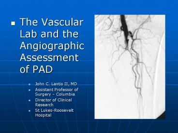
The Vascular Lab and the Angiographic Assessment of PAD PowerPoint PPT Presentation
1 / 21
Title: The Vascular Lab and the Angiographic Assessment of PAD
1
- The Vascular Lab and the Angiographic Assessment
of PAD - John C. Lantis II, MD
- Assistant Professor of Surgery Columbia
- Director of Clinical Research
- St Lukes-Roosevelt Hospital
2
The Questions
- Does the patient have enough blood flow to heal
their wound / or the intervention ? - Does the patient have PAD, and should I be
helping them to find coordinated care ? - Is the patients circulation compromised to the
point that I am highly concerned about tissue
loss ?
3
The Answers!
- (Obviously) A good physical exam
- Physiologic testing
- Ankle brachial index
- Pulse volume recording
- Duplex/MRI NOVA
- TCPO2 (Transcutaneous Oxygen Tension)
- Anatomic testing
- Duplex
- MRA
- Angiogram
- CTA
4
The Ankle Brachial Index
- Measurement of segmental leg pressure compared to
the highest brachial artery pressure - Can be done at the bedside
- Requires little equipment
- Helps determine level of disease
5
The ankle brachial Index
- Prognostic capabilities
- Forefoot amputations are likely to heal, if the
ankle pressure is gt 70 mmHg, or if the ABI gt 0.45 - Toe amputations are likely to heal with ankle
pressures of gt 35 mmHg or toe pressures gt 55 mmHg
- Limitations
- Ankle pressures can be artificially inflated in
patients with diabetes mellitus and ESRD - Toe pressures are therefore relied upon
- Pressure less than 50 mm Hg and a toe-to-arm
ratio of less than 0.6 is indicative of ischemic
arterial disease - Foot lesions usually heal if toe pressures exceed
30 mmHG in non-diabetic patients and 55 mmHG in
diabetic patients - Ipsilateral ankle to toe pressures can be used to
assess for obstructive pedal vascular disease - AVG 0.65 in normals
- AVG 0.23 in patients with rest pain of tissue loss
6
Pulse Volume Recordings
- More sensitive and more specific
- Probably the bread and butter physiologic test
- Will give good guidance to the level and severity
of disease
7
Pulse Volume Recordings(with ABI and exercise)
- Treadmill walking test
- Walking at 1.8 mph
- 10 incline
- Uncovers more subtle lesions
- Especially proximal lesions in the iliac and SFA
vessels - A fall in the ABI of 0.2 or a recovery to
baseline pressure that is greater than 1 minute
is significant
8
Categories of Chronic Limb Ischemia
- Clinical Description
- Normal Asymptomatic
- Mild Claudication
- (ABI - lt 0.7)
- Moderate Claudication
- Severe claudication
- Rest Pain
- (ABI - lt 0.4)
- Minor Tissue Loss
- Major Tissue Loss
- Pressure Criteria
- Normal Treadmill test
- Completes test, ankle pressure drops gt 20 mmHg,
absolute ankle pressure gt 50 mmHg - Between mild and severe
- Cannot complete treadmill test and ankle pressure
after exercise lt 50 mm Hg - Resting ankle pressure lt 60 mmHG or toe pressure
lt 40 mmHG - Resting ankle pressure less than 40 mmHg or toe
pressure less than 30 mmHg - Same as minor
9
Duplex Ultrasound(Combination of B mode imaging
and doppler velocity criteria)
- Doppler waveform analysis of the femoral,
popliteal and tibial vessels can be carried out - Waveforms are evaluated similarly to the PVR
tracings - More accurate at localizing disease than PVRs
- Very labor intensive
10
Transcutaneous Partial pressure of Oxygen
- Transcutaneous oxygen (tcPO2)
- Reflects the metabolic state of the target tissue
- Best for severe ischemia
- Heated Clark electrode (very tech dependent, hard
to reproduce) - lt 20 mmHg healing failure
- gt 40 mmHg healing success
- Elevate limb gt 300 /3 min drop gt 15 mmHg
healing failure
11
Other Methods of Assessing Blood Supply
- Laser Doppler Velocimetry
- A relative index of cutaneous blood flow
- With ischemia pulse waves are attenuated, mean
velocities are decreased - If mean velocity is gt 40 millivolts (mV) and
pulse wave amplitude is gt 4 mV associated with
healing - NOVA
- Non-invasive Optimal Vessel Analysis (NOVA) a
non-invasive Magnetic Resonance Imaging (MRI)
technique - NOVA provides actual milliliter/minute blood flow
data using specialized software analysis of
standard MRI phase contrast imaging - Investigational
12
Back to the Questions.
- Does the patient have enough blood flow to heal
their wound / or the intervention ? NO - Does the patient have PAD, and should I be
helping them to find coordinated care ? YES - Is the patients circulation compromised to the
point that I am highly concerned about tissue
loss ? YES
13
Leads to the next two questions
- Where is the patients lesion?
- Segmental Pressures
- Segmental PVRs
- Long leg duplex
- Can I get this patient revascularized?
- What type of lesion?
- How many and where?
14
MRA
- Non nephrotoxic contrast
- No arterial puncture
- However, claustrophobia limited
- Sensitivity and specificity to level of disease
80-85 - Approximately 85 concordance with Angiography
15
MRA
16
Angiography
- Usually nephrotoxic dye
- Arterial puncture
- Done with sedation (few issues with
claustrophobia) - Able to intervene at time of procedure
- With subtraction capabilities probably able to
see post-occluded vessels as well as MRA
17
Angiography
18
CT Angiogram
- Approaching MRAs capabilities
- Relatively large nephrotoxic dye load
- No arterial puncture
- Minimal claustrophobia issues
- Distal vessel resolution still machine and center
dependent
19
CT Angiogram
20
A day in the life.
- A patient limps in
- No palpable pulse
- Small amount of tissue loss
- ABI/PVRs are obtained
- .Obtain toe NIFs..
- Pt went onto heal..
21
Or more likely..
- We have flat line tracings
- Which we follow with a anatomic diagnostic .
- Which leads us to our next speakers

