Human Anatomy of the Urinary System PowerPoint PPT Presentation
Title: Human Anatomy of the Urinary System
1
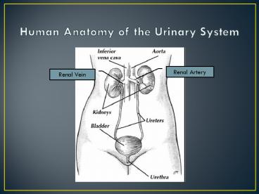
Human Anatomy of the Urinary System
Renal Artery
Renal Vein
2
Urinalysis
- Color of urine (clear and pale to deep yellow) is
due to urochrome which is a pigment that results
when hemoglobin is broken down - Some drugs and vitamins may alter urine color.
Cloudy urine is indicative of a urinary
infection.
3
Urine pH
- Usually slightly acidic (pH 6).
- Body metabolism and diet can cause urine pH to
vary from 4.5 to 8. - Large amounts of protein and whole wheat cause
acidic urine - Vegan diets or bacterial infections can cause pH
to climb and become alkaline (gt7)
4
Urine Specific Gravity
- SG is the ratio of the density of substance
divided by the density of water. - SG of water is 1.0, urine is 1.001-1.035
5
Chemical Composition
6
Abnormal components in urine
- Glucose condition is called Glycosuria. Caused
by diabetes mellitus or high intake of sugars - Proteins Proteinuria or Albuminuria. Caused by
pregnancy, high protein diets, heart failure,
severe hypertension, renal disease. - Hemoglobin Hemoglobinuria. Caused by
transfusion reaction or severe burns - Erythrocytes Hematuria. Cause by bleeding in
urinary tract (kidney stones). - Leukocytes Pyuria. Caused by urinary tract
infection.
7
Benedicts Test for Glucose
- Glucose is a reducing sugar- this means it has a
free aldehyde group - CuSO4 Cu SO4--
- 2 Cu Reducing Sugar Cu
(electron donor) - Cu Cu2O (ppt)
- The final color of the solution depends on how
much of this precipitate is formed and therefore
the color gives an indication of how much
reducing sugar was present. - Increasing amounts of reducing sugar
- green orange red brown
8
Biuret Test For Protein
- ? To about 2cm3 of test solution add an equal
volume of biuret solution, down the side of the
test tube. - ? A blue ring forms at the surface of the
solution, which disappears on shaking, and the
solution turns lilac-purple, indicating protein. - The colour change is due to a complex between
nitrogen atoms in a peptide chain and copper (II)
ions, so this is really a test for peptide bonds.
9
Purple (protein present)
PowerShow.com is a leading presentation sharing website. It has millions of presentations already uploaded and available with 1,000s more being uploaded by its users every day. Whatever your area of interest, here you’ll be able to find and view presentations you’ll love and possibly download. And, best of all, it is completely free and easy to use.
You might even have a presentation you’d like to share with others. If so, just upload it to PowerShow.com. We’ll convert it to an HTML5 slideshow that includes all the media types you’ve already added: audio, video, music, pictures, animations and transition effects. Then you can share it with your target audience as well as PowerShow.com’s millions of monthly visitors. And, again, it’s all free.
About the Developers
PowerShow.com is brought to you by CrystalGraphics, the award-winning developer and market-leading publisher of rich-media enhancement products for presentations. Our product offerings include millions of PowerPoint templates, diagrams, animated 3D characters and more.

