
LOWER LIMB DISSECTION PowerPoint PPT Presentation
1 / 91
Title: LOWER LIMB DISSECTION
1
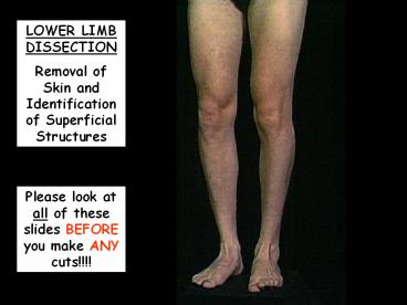
LOWER LIMB DISSECTION Removal of Skin and
Identification of Superficial Structures
Please look at all of these slides BEFORE you
make ANY cuts!!!!
2
- Make certain you know where the following
structures are located BEFORE you begin cutting.
Items in blue font on this slide are important
for presentation in BIOL 312 or BIOL 313. - great saphenous vein
- lesser saphenous vein
- accessory saphenous vein
- saphenous hiatus
- lateral femoral cutaneous nerve
- intermediate femoral cutaneous nerve
- medial femoral cutaneous nerve
- saphenous nerve
- cutaneous branches of the deep fibular nerve
- cutaneous branches of the superficial fibular
nerve
3
Begin with the cadaver in the supine position.
4
- Make an incision along the inguinal crease.
- Next, make a midline incision from the center
of cut 1 extending inferiorly over the center of
the patella, then over the anterior portion of
the leg, and over the center of the dorsum of the
foot.
5
- 3. Next make transverse circumferential
incisions at (a) the level of the infrapatellar
border, (b) just superior to the malleoli, and
(c) at the level of the metatarsal heads.
6
Then, reflect the skin from the midline,
taking care not to damage the superficial
structures (veins, and nerves) that lie
immediately deep to it.
7
For example, PLEASE try to save the great
saphenous vein. Note that it is contained WITHIN
much of the fat you will be removing.
8
Now that you have identified the path of the
great saphenous vein, remove the superficial
fascia and identify the deep fascia which
surrounds the muscles of the thigh and leg.
Remember that in the thigh, the deep fascia is
called the fascia latae. The fascia lata thickens
to form the strong iliotibial tract on the
lateral side of the thigh.
9
This is another view of the great saphenous
vein.
10
To find the great saphenous vein it is best to
first look distally. On the dorsum of the foot
you should see the dorsal venous arch. The two
largest superficial veins of the lower extremity
arise from it. On the medial side of the dorsal
venous arch, you should be able to identify the
great saphenous vein running anterior to the
medial malleolus.
11
On the medial side of the dorsal venous arch,
you should be able to identify the great
saphenous vein running anterior to the medial
malleolus.
12
On the lateral side of the dorsal venous arch,
you should be able to identify the lesser
saphenous vein running posterior to the lateral
malleolus.
lesser saphenous vein
dorsal venous arch
13
You should be able to identify the lesser
saphenous vein running posterior to the lateral
malleolus.
14
Now trace the great saphenous vein proximally.
15
Although the great saphenous vein is anterior to
the medial malleolus, it lies posterior to the
medial aspect of the knee, and then lies along
the medial aspect of the thigh.
16
As you continue to follow the great saphenous
vein proximally, you will see that approximately
5 to 10 cm below the inguinal ligament, this
vessel passes through an opening of the fascia
lata, called the saphenous hiatus to join with
the femoral vein.
17
You may see an accessory saphenous vein on the
medial aspect of the proximal thigh joining the
great saphenous vein before it enters the
saphenous hiatus.
18
This is another view of the accessory saphenous
vein on the medial aspect of the proximal thigh
joining the great saphenous vein before it enters
the saphenous hiatus.
19
You will see many perforating veins probe that
pierce the fascia lata and connect the
superficial veins to the network of deep veins of
the lower extremity.
20
At the region of the saphenous hiatus try to find
some inguinal lymph nodes. Some run parallel the
inguinal ligament and are generally located about
2 cm below it. Others are usually found along
both sides of the great saphenous vein near the
hiatus.
21
This is a view of some very enlarged lymph nodes
in the region of the saphenous hiatus.
22
Identify the lateral femoral cutaneous nerve
located over the upper lateral aspect of the
thigh.
23
Identify the intermediate femoral cutaneous nerve
located over the anterior aspect of the thigh.
Identify the medial femoral cutaneous nerve
located over the anterior aspect of the thigh.
24
Identify branches of the obturator nerve located
along the medial aspect of the superior thigh.
25
Identify the saphenous nerve located on the
anterior and medial aspect of the leg. This
nerve pierces the fascia lata superior to the
knee and then generally accompanies the great
saphenous vein on the medial aspect of the leg.
26
The saphenous nerve is located on the anterior
and medial aspect of the leg, where it generally
accompanies the great saphenous vein.
27
Now, return to the dorsum of the foot to look at
other cutaneous nerves.
28
Identify the cutaneous branches of the deep
fibular nerve between the first and second
metatarsals. (Obviously this cutaneous nerve
must convey sensation from a very small region.)
29
Identify the dorsal digital branches of the
superficial fibular nerve that provide cutaneous
innervation to most of the skin of the toes.
30
branches of superficial fibular nerve
deep fibular nerve
31
Turn the cadaver to the prone position and remove
the skin from the posterior aspect of the lower
extremities. This may be best accomplished by
making a vertical cut beginning at the
approximate level of the gluteal cleft, extending
inferiorly over the center of the popliteal
fossa, then over the posterior portion of the
leg, and over the center of the plantar surface
of the foot.
32
In this position you should be able to better
identify the lesser (small) saphenous vein as it
runs posterior to the lateral malleolus and onto
the posterior aspect of the leg.
33
(No Transcript)
34
- Make certain you know where the following
structures are located BEFORE you begin cutting.
Items in blue font on this slide are important
for presentation in BIOL 312 or BIOL 313. - femoral triangle
- femoral nerve
- femoral artery
- femoral vein
- great saphenous vein
- deep femoral artery (profunda femoris)
- medial femoral circumflex artery
- lateral femoral circumflex artery
- cutaneous branches of the superficial fibular
nerve
- Muscles
- Pectineus m.
- Iliopsoas m.
- Adductor longus m.
- Adductor magnus m.
- Adductor brevis m.
- Rectus femoris m.
- Vastus lateralis m.
- Vastus medialis m.
- Vastus intermedius m.
35
Muscles of the Anterior Compartment of the Thigh
36
- Rectus femoris muscle
- originates from the anterior inferior iliac spine
- only part of the quadriceps femoris muscle
- that crosses the hip joint
37
Vastus lateralis muscle
38
Vastus medialis muscle
39
Vastus intermedius muscle located deep to
portions of the other three parts of the
quadriceps femoris muscle so the rectus femoris
muscle must be retracted medially or laterally
40
superior inguinal ligament
lateral medial border of the sartorius muscle
medial lateral border of the adductor longus m.
Now that you have identified the muscles of the
quadriceps femoris, identify the boundaries and
contents of the femoral triangle.
41
inguinal ligament
42
Adductor longus m.
43
Sartorius m.
44
Adductor longus m.
Now that you have identified the borders of the
femoral triangle, identify the muscles that make
up its floor the rest of the adductor longus
m., the pectineus m., and the iliopsoas m.
45
Pectineus m.
46
Probe pointing to the iliopsoas m.
47
In the superior part of the femoral triangle, you
should see the great saphenous vein (from your
original superficial dissection) passing through
the saphenous hiatus to join the femoral vein.
48
Surrounding the femoral vein, femoral artery, and
associated deep inguinal lymph nodes is the
femoral sheath.
Great saphenous vein
49
Femoral vein
Femoral artery
Femoral nerve
Carefully open this femoral sheath and clean this
area so that we can clearly see the femoral vein,
the femoral artery, and the femoral nerve (from
medial to lateral V A N).
50
Muscular branches of the femoral n.
Now that you have a clear view of the femoral n.
in the femoral triangle, you should be able to
follow it distally to view its multiple muscular
branches (to the sartorius m., quadriceps femoris
m., and pectineus m. pectineus m. is also
innervated by a branch of the obturator n.).
You have already identified the cutaneous
branches of the femoral n. when you performed
your superficial dissection of this region
(intermediate and medial femoral cutaneous nn.).
The femoral n. continues as the saphenous n. in
the leg.
51
Now, look at some of the important branches of
the femoral artery.
52
The profunda femoris a. is the major branch of
the femoral a. in the femoral triangle. It
passes superficial to the pectineus m. and then
deep to the adductor longus m.. Along its course
it usually gives rise to four perforating
branches, which pass through openings in the
adductor magnus m. to supply the structures of
the posterior thigh.
53
Lateral circumflex a.
The circumflex aa. may be branches of either the
femoral a. or profunda femoris a.. They supply
blood to muscles in the region and participate in
collateral circulation at the hip joint. The
lateral circumflex a. leaves the femoral triangle
by passing deep to the sartorius m. and rectus
femoris m..
54
Medial circumflex a.
The medial circumflex a. usually leaves the
femoral triangle by traveling between the
adjoining borders of the iliopsoas and pectineus
mm.
55
If you look deep to the inferior aspect of the
sartorius m., you can identify the adductor
canal, which is the site where vessels pass from
the femoral triangle to the posterior aspect of
the knee (popliteal fossa). The adductor canal
begins at the apex of the femoral triangle and
ends, distally, at an opening in the tendon of
the adductor magnus muscle. This opening is
called the adductor hiatus.
Probe in adductor canal
56
So, the lateral wall of the adductor canal is
formed by the vastus medialis m.
57
And, the medial wall of the adductor canal is
made up of the adductor longus and adductor
magnus mm..
Adductor longus m.
Adductor magnus m.
58
Recall that we said that once the femoral nerve
gives off its muscular branches in the thigh, it
continues on with a name change. It is now
called the saphenous n.. This nerve accompanies
the femoral a. and v. in the adductor canal, but
it does not pass through the adductor hiatus.
59
On the medial side of the knee, the saphenous n.
passes anterior to the tendon of the adductor
magnus m. and pierces the deep fascia between the
tendons of the sartorius muscle and gracilis
muscle.
60
Muscles of the Medial Compartment of the Thigh
Gracilus m.
61
Probes indicate superior and inferior borders of
the adductor brevis m.
Although the adductor longus and pectineus mm.
are cut in this picture do NOT do this with
your dissection.
62
When observing the adductor brevis m., you should
also be able to view branches of the obturator n..
63
Adductor magnus m.
64
Now, lets look at structures in the leg.
65
Superior extensor retinaculum
Inferior extensor retinaculum
If you remove the deep (crural) fascia
surrounding the muscles of the leg, you can
identify the thickenings of the deep fascia that
hold the tendons of the muscles of the anterior
compartment of the leg in position at the ankle.
66
Now these retinacula must be cut to facilitate
identifying the tendons on the anterior aspect of
the ankle. It is best to identify these
structures at this site and then trace them
proximally to their bellies.
67
Tendon of tibialis anterior m.
68
Tendon of extensor hallucis longus m.
69
Deep fibular n.
70
Anterior tibial vessels
71
Tendons of extensor digitorum longus m.
72
Tendon of fibularis tertius m.
73
Now that you have identified the structures at
the anterior ankle, trace the tendons proximally
to identify muscle bellies in the anterior
compartment of the leg.
74
Tibialis anterior m.
Extensor digitorum longus m.
75
Extensor hallucis longus m.
Extensor digitorum longus m.
76
Fibularis tertius m. arising from tendons of
extensor digitorum longus tendons
77
Now identify the structures of the lateral
compartment of the leg.
78
Fibularis longus m. remember that the tendon
of this muscle travels on the plantar aspect of
the foot and inserts on the medial cuneiform and
first metatarsal.
79
If the fibularis longus m. is retracted at its
most proximal site, you should be able to observe
these two nerves.
Superficial fibular n.
Deep fibular n.
80
Fibularis brevis m. note its insertion onto
the base of the fifth metatarsal.
81
Place the cadaver in the prone position to look
at the structures in the posterior compartment of
the leg.
82
Medial head of the gastrocnemius m.
Lateral head of the gastrocnemius m.
83
To best observe the other muscles of this
compartment, the two heads of the gastrocnemius
m. need to be cut and reflected. However, do
NOT do this until you have been told to do so.
84
To best observe the other muscles of this
compartment, the two heads of the gastrocnemius
m. need to be cut and reflected. However, do
NOT do this until you have been told to do so.
85
Soleus m.
86
Popliteus m.
87
Plantaris m.
tendon of plantaris m. inserting into
medial border of calcaneal tendon
88
Now, to best look at the other muscles we would
like to identify, the soleus muscle needs to be
cut and reflected. However, once again, do NOT
do this until you have been specifically told to
do so.
89
Observe the tendons of these muscles at the level
of the ankle.
Flexor digitorum longus m.
Tibialis anterior m.
90
Flexor hallucis longus m.
Observe the tendons of these muscles at the level
of the ankle.
91
Tibial n. and posterior tibial vessels
Recommended
