Microscopes - PowerPoint PPT Presentation
1 / 40
Title: Microscopes
1
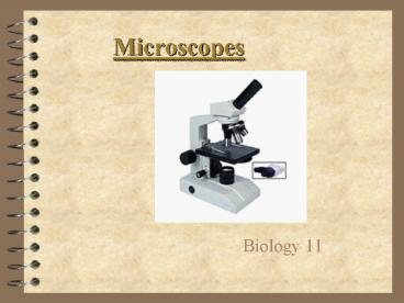
Microscopes
- Biology 11
2
The History
- Many people experimented with making microscopes
- Was the microscope originally made by accident?
(Most people were creating telescopes) - The first microscope was 6 feet long!!!
- The Greeks Romans used lenses to magnify
objects over 1000 years ago.
3
The History
- Hans and Zacharias Janssen of Holland in the
1590s created the first compound microscope
- Zacharias Jansen
- 1588-1631
4
The History
- Anthony van Leeuwenhoek and Robert Hooke made
improvements by working on improving the lenses
Robert Hooke 1635-1703
Anthony van Leeuwenhoek 1632-1723
Hooke Microscope
5
How a Microscope Works
Convex Lenses are curved glass used to make
microscopes (and glasses etc.)
Convex lenses bend light and focus it in one spot.
6
How a Microscope Works
Objective Lens (Gathers Light, Magnifies And
Focuses Image Inside Body Tube)
Ocular Lens (Magnifies Image)
Body Tube (Image Focuses)
- Bending Light The objective (bottom) convex lens
magnifies and focuses (bends) the image inside
the body tube and the ocular convex (top) lens
of a microscope magnifies it (again).
7
The Parts of a Microscope
8
Ocular Lens
Body Tube
Nose Piece
Arm
Objective Lenses
Stage
Stage Clips
Coarse Adjustment
Diaphragm
Fine Adjustment
Light Source
Base
9
Body Tube
- The body tube holds the objective lenses and the
ocular lens the proper distance apart
Diagram
10
Nose Piece
- The Nose Piece holds the objective lenses and can
be turned to choose a different magnification
objective.
Diagram
11
Objective Lenses
- The Objective Lenses increase magnification
(usually from 4x to 40x)
Diagram
12
Stage Clips
- These 2 clips hold the slide/specimen in place on
the stage.
Diagram
13
Diaphragm
- The Diaphragm controls the amount of light on the
slide/specimen
Turn to let more light in or to make dimmer.
Diagram
14
Light Source
- Projects light upwards through the diaphragm, the
specimen and the lenses - Some have lights, others have mirrors where you
must move the mirror to reflect light
Diagram
15
Ocular Lens/Eyepiece
- Magnifies the specimen image
Diagram
16
Arm
- Used to support the microscope when carried.
Holds the body tube, nose piece and objective
lenses
Diagram
17
Stage
- Supports the slide/specimen
Diagram
18
Coarse Adjustment Knob
- Moves the stage up and down (quickly) for
focusing your image, use only with low power
objective lens!
Diagram
19
Fine Adjustment Knob
- This knob moves the stage SLIGHTLY to sharpen the
image
Diagram
20
Base
- Supports the microscope
Diagram
21
Label the microscope diagram
- Page 627 in the textbook will help
22
Parts of a microscope
23
What the parts do
- the lens you look through, magnifies the specimen
- supports the microscope
- holds objective lenses
- magnify the specimen (2)
- supports upper parts of the microscope, used to
carry microscope - used to focus when using the high power objective
- where the slide is placed
- regulates the amount of light reaching the object
- used to focus when using the low power objective
- provides light
- hold slide in place on the stage
24
Caring for a Microscope
- Carry it with 2 HANDSone on the arm and the
other on the base. - Make sure its on a flat surface, away from the
edge of the desk. - Dont pull on the cord to unplug it.
- Clean lenses only with lens tissue.
25
Carry a Microscope Correctly
26
Using a Microscope
- Start on the lowest magnification, with the stage
lowered. - Place slide on stage and lock with clips.
- Use coarse adjustment knob to center the sample
and focus on the object. - Adjust light source using the diaphragm.
- Use fine adjustment to focus.
27
Using a Microscope (contd)
- Rotate the nosepiece to increase magnification
from lowest to highest. - Use only the fine adjustment knob to focus on
high power.
28
Microscopy involves 2 processes
- Magnification
- enlargement
- Resolution (resolving power)
- - sharpness of the image
29
Magnification
- The ability to increase the size of the image of
a specimen. - Total magnification of an image is found by
multiplying the power of the ocular lens by the
power of the objective. - The larger the magnification the less area you
are able to see.
30
Resolution
- The ability to see details clearly.
- in order for the increased magnification of a
microscope to be of use, its resolution must also
be increased - resolution is dependant on the quality of the
lens and is limited when using light microscopes
31
Microscope video.
- Fill out response while you watch.
32
Simple Microscope
- Anton van Leewenhoek was the first to use the
single lens microscope for biological purposes. - Consisted of a single lens
- used today for quick observations in the field
- specimens may be alive for examination.
- but it has low magnification
33
Compound Light Microscope
- Consists of two lenses, the ocular and the
objective, both of which magnify the image - these two lenses form the optical system.
- Structural parts that hold the specimen and the
lenses is called the mechanical system
34
Compound Light Microscope
- Uses
- good for basic lab work (up to 1500x)
- can be used to view living things but most
specimens are dead - usually used with stain
- Disadvantages
- cannot view cell structures
- low resolution
35
Stereo microscope (dissection)
- Two sets of lenses- an ocular and an objective
for each eye - Uses
- allows the scientist to view images as 3-D
- used to study external structures and for
dissections - specimens may be kept alive for examination.
- Disadvantage
- Low magnification (6-50x)
36
Phase Contrast (lens) Microscope
- Changes the way light passes through a living
specimen with phase plates - Can see cell structures that are not usually
visible under a compound microscope. - Useful for examining live specimens
- But has a low magnification
Frits (Frederik) Zernike won the Nobel prize 1953
for the development of this microscope.
37
Transmission Electron Microscope
- Electron beam is sent through a vacuum chamber
then electromagnets focus the image on a screen - an image can be magnified a million times so you
can see cell structures and specimens smaller
than a cell - Vacuum is needed, specimens are dead and required
complex procedures to prepare them
Also know as the TEM
38
Scanning Electron Microscope
- Like the TEM, this microscope uses electrons to
focus an image and is able to view things much
smaller than the light microscope. - Unlike the TEM, the SEM allows the viewing of 3D
images and has better resolution. - Vacuum is needed, specimens are dead and required
complex procedures to prepare them. Lower
magnification possible
Also known as the SEM
39
Scanning electron micrograph (SEM) of various
Pollen. Public domain image reference Dartmouth
Electron Microscope Facility, Dartmouth College
40
Microscope Calculations


