Structures and Functions of the Respiratory System - PowerPoint PPT Presentation
1 / 63
Title:
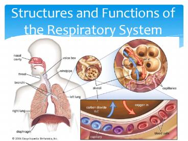
Structures and Functions of the Respiratory System
Description:
Title: Alterations in Respiratory Function Author: Evelyn Last modified by: Ramon Created Date: 8/16/2006 12:00:00 AM Document presentation format – PowerPoint PPT presentation
Number of Views:232
Avg rating:3.0/5.0
Title: Structures and Functions of the Respiratory System
1
Structures and Functions of the Respiratory System
2
Gas Exchange
- Ventilation
- Diffusion (alveolar-capillary membrane)
- Perfusion
- Diffusion (capillary-cellular level)
3
Ventilation Movement of Chest Wall
4
Ventilation
- Depends on volume and pressure changes within
thoracic cavity - Diaphragm is major muscle of inspiration also
external intercostal muscles. Contraction
increases diameter of thoracic cavity? ?
intrathoracic pressure ?air flows into
respiratory system - Expiration is passive process d/t lung
elasticity. ? intrathoracic pressure? air flows
out of lungs - Accessory muscles
5
Control of Ventilation
- Neural control- respiratory center in medulla
pons - Central chemoreceptors sensitive to pH
- Peripheral chemoreceptors- sensitive to paO2
- Patients with COPD- hypoxic drive
- WOB- amount of effort required for the
maintenance of a given level of ventilation (as
WOB ?s, more energy is expended for adequate
ventilation)
6
Factors Influencing Ventilation
- Airway resistance- opposition to gas flow
- Compliance- distensibility / stretchability
- - Dependent on lung elasticity elastic
recoil - of chest wall
- - Decreased compliance- lungs difficult to
- inflate
- - Increased compliance- destruction of
- alveolar walls loss of tissue elasticity
7
DiffusionAlveolar-Capillary Membrane
8
Oxyhemoglobin Curve
9
Ventilation-Perfusion
- Adequate diffusion depends on balanced
ventilation-perfusion (V/Q) ratio - Normal lung V4L/min Q 5L/min (0.8)
- If imbalanced gas exchange interrupted
- - High V/Q wasted or dead-space
- ventilation
- - Low V/Q blood shunted past area no
- gas exchange occurs
10
V/Q Matching
11
Perfusion
12
DiffusionBody Tissue-Blood Capillary
13
COPD
- Progressive, irreversible airflow limitation
- Associated with abnormal inflammatory response of
lungs to noxious particles or gases
14
COPDEtiology
- Cigarette smoking
- Occupational chemicals and dusts
- Air pollution
- Infection
- Heredity- A1-antitrysin deficiency
- Aging
15
COPDPathophysiology
- Primary process is inflammation
- Inhalation of noxious particles? inflammatory
cells release mediators (leukotrienes,
interleukins, TNF) ? airways become inflammed
with increased goblet cells ? excess mucus
production (bronchitis) structural remodeling
to peripheral airways with ?d collagen scar
tissue
16
COPDPathophysiology
- Destruction of lung tissue caused by imbalance of
proteinases/antiproteinases results in emphysema
with loss of attachments peripheral airway
collapse (Centrilobar- affects respiratory
bronchioles/upper lobes/mild disease panlobar-
alveolar ducts, sacs, respiratory bronchioles-
lower lobes/AAT deficiency
17
COPDPathophysiology
- Air goes into lungs easily but unable to come
out air trapped in distal alveoli, causing
hyperinflation overdistension - PV thickens with ?surface area for gas exchange-
V/Q mismatch
18
(No Transcript)
19
COPDChronic Bronchitis vs. Emphysema
20
Emphysema
21
Chronic BronchitisBlue Bloater versus Pink Puffer
22
COPD Behaviors
- Develop slowly around 50 years of age after
history of smoking - Cough, sputum production, dyspnea
- In late stages, dyspnea at rest
- Wheezing/chest tightness- may vary
- Prolonged IE, ?BS, tripod position, pursed-lip
breathing, edema - ? A-P diameter of chest
- Advanced- weight loss, anorexia (hypermetabolic
state) - Hypoxemia, possible hypercapnia
- Bluish-red color from polycythemia, cyanosis
23
Increased A-P DiameterBarrel-Chest
24
COPDDiagnosis
- PFTs (? RV, ?FEV1)
- CXR
- ABGs
- Sputum CS if infection suspected
- EKG- RV hypertrophy
- 6 minute oxy-walk
25
(No Transcript)
26
COPD Classification
Spirometry Results
Stage I Mild FEV1/FVC lt 0.70 FEV1 80 predicted
Stage II Moderate FEV1/FVC lt 0.70 50 FEV1 lt 80 predicted
Stage III Severe FEV1/FVC lt 0.70 30 FEV1 lt 50 predicted
Stage IV Very Severe FEV1/FVC lt 0.70 FEV1 lt 30 predicted OR FEV1 lt 50 predicted PLUS chronic respiratory failure
27
COPDComplications
- Cor pulmonale- RV hypertrophy 2º pulmonary
hypertension (late) - Exacerbations of COPD
- Acute respiratory failure
- Peptic ulcer and gastroesophageal reflux disease
- Depression/anxiety
28
COPD- Collaborative Care
- Smoking cessation
- Medications- bronchodilators (inhaled
step-wise), Spriva (LA anticholinergic), ICS - Oxygen therapy
- RT- PLB, diphragmatic, cough, CPT, nebulization
therapy - Nutrition- Avoid over/underweight, rest 30
before eating, 6 small meals, avoid foods that
need a great deal of chewing, avoid exercise 1 hr
before meal, take fluids between meals to avoid
stomach distension
29
COPDNursing Diagnoses
- Ineffective Breathing Pattern
- Impaired Gas Exchange
- Ineffective Airway Clearance
- Imbalanced Nutrition Less than Body Requirements
30
Asthma
- Chronic inflammatory disorder associated with
airway hyperresponsiveness leading to recurrent
episodes (attacks) - Often reversible airflow limitation
- Prevalence increasing in many countries,
especially in children
31
AsthmaPathophysiology
- Airway hyperresponsiveness as a result of
inflammatory process - Airflow limitation leads to hyperventilation
- Decreased perfusion ventilation of alveoli
leads to V/Q mismatch - Untreated inflammation can lead to LT damage that
is irreversible - Chronic inflammation results in airway remodeling
32
(No Transcript)
33
(No Transcript)
34
AsthmaPotential Triggers
- Allergens 40
- Exercise (EIA)
- Air pollutants
- Occupational factors
- Respiratory infections viral
- Chronic sinus and nose problems
- Drugs and food additives ASA, NSAIDs,
ß-blockers, ACEi, dye, sulfiting agents - Gastroesophageal reflux disease (GERD)
- Psychological factors- stress
35
Asthma Inflammation Effects
- Bronchospasm
- Plasma exudation
- Mucus secretion
- AHR
- Structural changes
36
Asthma InflammationClinical Manifestations
- Cough
- Chest tightness
- Wheeze
- Dyspnea
- Expiration prolonged -13 or 14, due to
bronchospasm, edema, and mucus - Feeling of suffocation- upright or slightly bent
forward using accessory muscles - Behaviors of hypoxemia- restlessness, anxiety,
?HR BP, PP
37
AsthmaDiagnosis
- History and patterns of symptoms
- Measurements of lung function
- PFTs- usually WNL between attacks ? FVC, FEV1
- PEFR- correlates with FEV
- Measurement of airway responsiveness
- CXR
- ABGs
- Allergy testing (skin, IgE)
38
AsthmaTherapeutic Goals
- No (or minimal) daytime symptoms
- No limitations of activity
- No nocturnal symptoms
- No (or minimal) need for rescue medication
- Avoid adverse effects from asthma medications
- Normal lung function
- No exacerbation
- Prevent asthma mortality
- Minimal twice or less per week
39
AsthmaCollaborative Management
- Suppress inflammation
- Reverse inflammation
- Treat bronchoconstriction
- Stop exposure to risk factors that sensitized the
airway
40
AsthmaMedications
- Antiinflammatory Agents
- Corticosteroids- suppress inflammatory response.
Reduce bronchial hyperresponsiveness mucus
production, ? B2 receptors - Inhaled preferred route to minimize systemic
side effects - Teaching
- Monitor for oral candidiasis
- Systemic many systemic effects monitor blood
glucose - Mast cell stabilizers- NSAID inhibit release of
mediators from mast cells suppress other
inflammatory cells (Intal, Tilade)
41
AsthmaMedications
- Antiinflammatory Agents
- Leukotriene modifiers
- Block action of leukotrienes
- Accolate, Singulair, Zyflo)
- Not for acute asthma attacks
- Monclonal Ab to IgE
- ? circulating IgE
- Prevents IgE from attaching to mast cells, thus
preventing the release of chemical mediators - For asthma not controlled by corticosteroids
- Xolair SQ
42
AsthmaMedications
- Bronchodilators
- B-agonists- SA for acute bronchospasm to
prevent exercised induced asthma (EIA)
(Proventil, Alupent) LA for LT control - Combination ICS LA B-agonist (Advair)
- Methylxanthines- Theophylline alternative
bronchodilator if other agents ineffective.
Narrow margin of safety high incidence of
interaction with other medications - Anticholinergics- block bronchoconstriction .
Additive effect with B-agonists (Atrovent)
43
AsthmaPatient Teaching- Medications
- Name/dosage/route/schedule/purpose/SE
- Majority administered by inhalation (MDI, DPI,
nebulizers) - Spacer MDI- for poor coordination
- Care of MDI- rinse with warm H2O 2x/week
- Potential for overuse
- Poor adherence with asthma therapy is challenge
for LT management - Avoid OTC medications
44
AsthmaCollaborative Care
- GINA- decrease asthma morbidity/mortality
improve the management of asthma worldwide - Education is cornerstone
- Mild Intermittent/Persistent avoid triggers,
premedicate before exercise, SA or LA Beta
agonists, ICS, leukotriene blockers - Acute episode Oxygen to keep O2Satgt90, ABGs,
MDI B-agonist if severe- anticholinergic
nebulized w/B agonist, systemic corticosteroids
45
AsthmaNursing Diagnoses
- Ineffective Airway Clearance
- Impaired Gas Exchange
- Anxiety
- Deficient Knowledge
46
Pneumonia
- HAP- pneumonia occurring 48 hours or longer after
admission - VAP- pneumonia occurring 48-72 hours after ET
intubation - HCAP- hospitalized for 2 or more days within 90
days of infection resided in LTC facility
received IV therapy or wound care within past 30
days of current infection attended a hospital or
dialysis clinic - Aspiration pneumonia- abnormal entry of
secretions into lower airway
47
PneumoniaPathophysiology
- Congestion
- Fluid enters alveoli organisms multiply
infection spreads - Red hapatization
- Massive capillary vasodilation alveoli filled
with organisms, neutrophils, RBCs, fibrin - Gray hepatization
- Blood flow decreases leukocytes fibrin
consolidate in affected part - Resolution
- Resolution healing exudate processed by
macrophages
48
PneumoniaRisk Factors
- Aging
- Air pollution
- Altered LOC
- Altered oral normal flora secondary to
antibiotics - Prolonged immobility
- Chronic diseases
- Debilitating illness
- Immunocompromised state
- Inhalation or aspiration of noxious substances
- NG tube feedings
- Malnutrition
- Resident of Long-term care
- Smoking
- Tracheal intubation
- Upper respiratory tract infection
49
Pneumonia Behaviors
- Usually sudden onset
- Fever, shaking chills, SOB, cough w/purulent
sputum, pleuritic CP - Elderly/debilitated- confusion or stupor
50
Pneumonia- Complications
- Pleuritis
- Pleural effusion- 40 of hospitalized patients
- Atelectasis
- Bacteremia
- Lung abscess
- Empyema
- Pericarditis
51
PneumoniaDiagnostic Studies
- CXR
- Sputum CS
- Blood cultures
- ABGs
- Leukocytosis
52
Pleural Effusion
53
Pneumonia
54
PneumoniaCollaborative Care
- Prompt treatment with antibiotics
- Oxygen, analgesics, antipyretics
- Influenza vaccine
- Pneumococcal vaccine
- Nutrition
- PSI Pneumonia Patient Outcomes Research Team
Severity Index - Determine whether to treat at home or in hospital
55
PneumoniaNursing Assessment
- Fever in any hospitalized patient
- Pain
- Tachypnea
- Use of accessory muscles
- Rapid, bounding pulse
- Relative bradycardia
- Coughing
- Purulent sputum
56
PneumoniaNursing Assessment
- Consolidation
- Auscultation
- Bronchial breathing
- Bronchovesicular rhonchi
- Crackles
- Fremetis
- Egophony
- Whispered pectroloquy
57
PneumoniaNursing Diagnoses
- Ineffective airway clearance RT copious
tracheobronchial secretions - Activity intolerance RT altered respiratory
function - Risk for fluid volume deficit RT fever and
dyspnea - Knowledge deficit about the treatment regimen and
preventive health measures
58
PneumoniaPotential Problems
- Hypotension and shock
- Respiratory failure
- Atelectasis
- Pleural effusion
- Delerium
- Superinfection
59
PneumoniaNursing Goals
- Improving airway patency
- Conserving energy rest
- Maintaining proper fluid balance
- Patient understanding of treatment and prevention
- Prevention of complications
60
PneumoniaNursing Interventions
- Improving airway patency
- Removing secretions coughing vs. suctioning
- Adequate hydration loosens secretions
- Air humidification to loosen secretions and
improve ventilation - Chest physiotherapy loosens and mobilizes
secretions
61
PneumoniaNursing Interventions
- Promoting rest and conserving energy
- Bedrest with frequent changes of position
- Energy conservation
- Sedatives to decrease work of breathing and
energy expenditure unless contraindicated - Promoting fluid intake
- Dehydration is possible RT insensible fluid
losses through respiratory tract - If not contraindicated, increase fluid intake to
2 liters/day
62
PneumoniaNursing Interventions
- Patient education and home care considerations
- Increase activities as tolerated fatigue and
weakness may be prolonged - Breathing exercises to clear the lungs should be
taught - Smoking cessation if indicated smoking destroys
tracheobronchial ciliary action, which is the
first line of defense for the lungs. Smoking
also irritates the mucus cells of the bronchi and
inhibits the function of alvolar macrophages - Patient is encouraged to get influenza vaccine
because influenza increases risk for secondary
bacterial infections - Staphylococcus
- H. influenzae
- S. pneumonae
- Encouraged to get Pneumovax against S. pneumonae
63
Pneumonia- Core Measures
- Oxygenation assessment (ABGs, oximetry)
- Pneumococcal vaccine (gt65yo prior to DC)
- BC performed within 24h prior to after hospital
arrival - BC before first antibiotic
- Adult smoking cessation advice
- Antibiotic timing- within 4 hours of arriving to
hospital - Influenza vaccine


