
Background of Anatomy and Physiology - PowerPoint PPT Presentation
1 / 80
Title:
Background of Anatomy and Physiology
Description:
Background of Anatomy and Physiology Human skeleton made up of 206 bones 1. Axial skeleton includes a. Bones of skull b. Ribs and sternum c. Vertebral column – PowerPoint PPT presentation
Number of Views:105
Avg rating:3.0/5.0
Title: Background of Anatomy and Physiology
1
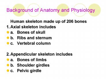
Background of Anatomy and Physiology
- Human skeleton made up of 206 bones
- 1. Axial skeleton includes
- a. Bones of skull
- b. Ribs and sternum
- c. Vertebral column
- 2. Appendicular skeleton includes
- a. Bones of limbs
- b. Shoulder girdles
- c. Pelvic girdle
2
Classification of bones by shape
3
Functions of bones
- 1. Form structure and provide support for soft
tissues - 2. Protect vital organs from injury
- 3. Serve to move body parts by providing points
of attachment for muscles - 4. Store minerals
- 5. Serve as site for hematopoiesis
- Bone cells include
- 1. Osteoblasts cells that form bone
- 2. Osteocytes cells that maintain bone matrix
- 3. Osteoclasts cells that resorb bone
4
Clients with Musculoskeletal Disorders
- Background
- 1. Normal bone remodeling process involves
sequence of bone reabsorption and formation - 2. Adults replace about 25 of trabecular bone
(the porous type of bone found in the spine and
all articulating joints) every 4 months through
reabsorption of old bone by osteoclasts and
formation of new bone by osteoblasts
5
Client with osteoporosis
- Definition
- a. Disorder characterized by loss of bone mass,
increased bone fragility, increased risk for
fractures - b. Imbalance of processes that influence bone
growth and maintenance associated with aging,
but may result from endocrine disorder or
malignancy - c. Significant health threat for Americans
estimated 28 million persons more common in
aging women half of women over 50 experience
osteoporosis-related fracture in lifetime (hip,
wrist, vertebrae)
6
Client with osteoporosis
- Risk Factors
- a. Risk of developing osteoporosis depends on
amount of bone mass achieved between ages 25 35 - b. Unmodifiable risk factors
- 1. Aging decrease in osteoblastic and
osteoclastic activity related to decreasing
levels of hormones (estrogen in females
testosterone in males) - 2. Gender women have 10 15 less peak bone
mass than men bone loss begins earlier (30s)
and proceeds more rapidly (before menopause) - 3. European Americans and Asians have less bone
density than African Americans - 4. Endocrine disorders affecting metabolism
hyperthyroidism, hyperparathyroidism, Cushings
syndrome, diabetes mellitus
7
Client with osteoporosis
- Modifiable risk factors
- 1. Calcium deficiency insufficient calcium in
diet results in body removing calcium from bones
diets high in protein lead to acidosis, and high
in diet soda are high in phosphate - 2. Menopause, decreasing estrogen levels
estrogen replacement therapy can reverse bone
changes but may increase risk for other diseases - 3. Cigarette smoking decreased blood supply to
bones - 4. Excessive alcohol intake toxic effect on
osteoblastic activity high alcohol intake
frequently associated with nutritional
deficiencies - 5. Sedentary life style weight-bearing exercise
such as walking positively influences bone
metabolism - 6. Use of specific medications
aluminum-containing antacids, corticosteroids,
anticonvulsants, prolonged heparin therapy,
antiretroviral
8
A normal spine at 40 years, and the osteoporotic
changes at ages 60 and 70 years
9
Client with osteoporosis
- Pathophysiology
- a. Diameter of bone increases, thinning outer
supportive cortex - b. Trabeculae (spongy tissue) lost and outer
cortex thins - c. Minimal stress leads to fracture
- 4. Manifestations (silent disease bone loss
occurs without symptoms) - a. Loss of height
- b. Progressive curvature of spine (dorsal
kyphosis, cervical lordosis, accounting for
dowagers hump) - c. Low back pain
- d. Fractures of forearm, spine or hip
10
Client with osteoporosis
- Complications
- a. Fractures (gt 1.5 million fractures yearly),
many spontaneous or resulting from everyday
activities - b. Persistent pain and associated posture changes
restrict client activities and ability to perform
ADL - 6. Collaborative Care
- a. Stopping or slowing osteoporosis
- b. Alleviating symptoms
- c. Preventing complications
11
Client with osteoporosis
- Diagnostic Tests
- a. Xrays picture of skeletal structures but
osteoporotic changes not seen untilgt 30 of bone
mass lost - b. Quantitative computed tomography (QCT) of
spine measures trabecular bone within vertebral
bodies - c. Dual-energy Xray absorptiometry (DEXA)
measures bone density in lumbar spine or hip
highly accurate - d. Alkaline phosphatase (AST) elevated post
fracture - e. Serum bone Gla-protein (osteocalcin) marker of
osteoclastic activity and is indicator of rate of
bone turnover used to evaluate effects of
treatment
12
Client with osteoporosis
- Medications
- a. Estrogen replacement therapy reduces bone
loss, increases bone density in spine and hip,
reducing risk of fractures in postmenopausal
women. - 1. Recommended for women who have undergone
surgical menopause before age 50 - 2. Associated risk for estrogen therapy alone is
increased risk of endometrial cancer - 3. Hormone replacement therapy (estrogen and
progestin) associated with increased risk for
cardiovascular disease and breast cancer - b. Raloxifene (Evista) selective estrogen
receptor modulator (SERM) that prevents bone loss
by mimicking estrogen effects on bone density
side effects are hot flashes contraindicated for
women with history of blood clots - c. Biphosphonates potent inhibitors of bone
resorption used to prevent and treat osteoporosis - 1.Alendronate (Fosamax)
- 2.Risedronate (Actonel)
- 3.Etidronate (Didronel)
- d. Calcitonin (Miacalcin) hormone increases bone
formation and decreases bone resorption
available as nasal spray or parenteral - e. Sodium fluoride stimulates osteoblast
activity, decreases risk of spinal fractures but
associated with increased risk of other fractures
including hip
13
Client with osteoporosis
- Nursing Care
- a. Emphasis is prevention and education of
clients under age of 35 - b. Prevention of complications in those with
osteoporosis - Health Promotion
- a. Calcium intake
- 1. Maintain daily intake of calcium at
recommended levels, in divided doses - a. Age 19 50 1000mg
- b. Age 51-64 1200 mg
- c. Age 65 and gt 1500 mg)
- 2. Optimal intake before age 30 35 increases
peak bone mass - 3. Foods high in calcium include milk, milk
products, salmon, sardines, clams, oysters, dark
green leafy vegetables - 4. Supplementationcalcium carbonate (Tums)
calcium combined with Vitamin D for older adults
14
Client with osteoporosis
- Exercise
- 1. Physical activity that is weight-bearing
- 2. Walking 20 minutes, 4 or gt times per week
- Health-related behaviors
- 1. Include not smoking
- 2. Avoid excessive alcohol
- 3. Limit caffeine to 2 3 cups of coffee daily
- 4. Limit diet soda
15
Client with osteoporosis
- Nursing Diagnoses
- a. Health Seeking Behaviors
- b. Risk for Injury
- c. Imbalanced Nutrition Less than body
requirements - d. Acute Pain
- Home Care Focus is on education including safety
and fall prevention inside and outside the home
16
Client with Pagets Disease (osteitis deformans)
- Description
- a. Progressive skeletal disorder with excessive
metabolic bone activity leading to affected bones
becoming larger and softer - b. Affects femur, pelvis, vertebrae, sacrum,
sternum, skull - c. Relatively rare
- d. Occurs more often in whites
- e. Slightly more common in males
- f. Familial tendency
17
Client with Pagets Disease (osteitis deformans)
- Pathophysiology
- a. Bones are initially soft and bowing occurs
then become hard and brittle leading to fractures
- b. Slow progression with 2-stage process
- 1. Excessive osteoclastic bone resorption
- 2. Excessive osteoblasticbone formation
18
Client with Pagets Disease (osteitis deformans)
- Manifestations
- a. Most are asymptomatic
- b. Localized pain of long bones, spine, pelvis,
cranium pain is mild to moderate deep ache which
is aggravated by pressure and weight-bearing
noticed at night and when resting - c. Flushing and warmth over areas of bone
involvement
19
Client with Pagets Disease (osteitis deformans)
- Complications
- a. Degenerative osteoarthritis
- b. Pathological fractures
- c. Nerve palsy syndromes from involvement of
upper extremities - d. Compression of spinal cord causing tetraplegia
- e. Mental deterioration from skull involvement
and brain compression
20
Client with Pagets Disease (osteitis deformans)
- Collaborative Care
- a. Pain relief
- b. Suppression of bone cell activity
- c. Complication prevention
- Diagnostic Test
- a. Xray (often incidental) slow localized areas
of demineralization in early phase later
enlargement of bones with tiny cracks in long
bones or bowing in weight-bearing bones - b. Bone scan active Pagets disease
21
Client with Pagets Disease (osteitis deformans)
- c. CT scans and MRI show degenerative problems,
spinal stenosis, nerve root impingement - d. Serum alkaline phosphatase steady rise as
disease progresses - e. Urinary collagen pyridinoline testing
indicator of rate of bone resorption
22
Client with Pagets Disease (osteitis deformans)
- Medications
- a. Mild symptoms relieved by aspirin or NSAIDs
- b. Bone resorption retarded by
- 1. Biphosphonates calcium supplements are
prescribed in addition - a. Alendronate (Fosamax)
- b. Pamidronate (Aredia)
- c. Tiludronate (Skelid)
- 2. Calcitonic works as analgesic for bone pain
- a. Salmon calcitonin (Calcimar)
- b. Human calcitonin (Cibacalcin)
23
Client with Pagets Disease (osteitis deformans)
- Surgery
- a. Total hip or knee replacement is usually
required when client with Pagets disease
develops degenerative arthritis of hip or knee - b. May require surgery for spinal stenosis, nerve
root compression - Nursing Diagnoses
- a. Chronic Pain
- 1. May involve wearing a back brace for relief of
back pain - 2. Heat therapy and massage
- b. Impaired Physical Mobility
- Home Care manifestations often relieved by
treatment
24
Client with osteomalacia (adult rickets)
- Metabolic bone disorder characterized by
inadequate or delayed mineralization of bone
matrix leading to marked deformities of weight
bearing bone and pathologic fractures - Pathophysiology
- a. Primary causes are vitamin D deficiency and
hypophosphatemia - 1. Vitamin D deficiency
- a. Present in
- 1. Older adults
- 2. Very-low-birth weight infants
- 3. Strict vegetarians
- b. Caused by
- 1. Diet low in vitamin D
- 2. Impaired intestinal absorption of fats
- 3. Inadequate sun exposure
- 4. Some types of renal failure
- 2. Hypophosphatemia most commonly caused by
alcohol abuse
25
Process of vitamin D metabolism in the body
26
Client with osteomalacia (adult rickets)
- Other causes
- 1. Insufficient calcium absorption in intestines,
due to lack of calcium or resistance to action of
Vitamin D - 2. Increase loss of phosphorus through urine
- Manifestations
- a. Bone pain and tenderness
- b. Common fractures are distal radius and
proximal femur - Collaborative Care requires differential
diagnosis from osteoporosis
27
Client with osteomalacia (adult rickets)
- Diagnostic Tests
- a. Xray demonstrates generalized bone
demineralization - b. Serum calcium levels are normal or low
- c. Serum parathyroid hormone is frequently
elevated as compensatory response - d. Alkaline phosphatase level usually elevated
28
Client with osteomalacia (adult rickets)
- Medications
- a. Treatment of underlying condition
- b. Vitamin D therapy with calcium and phosphate
supplements - c. Radiologic evidence of healing apparent within
weeks of therapy
29
Client with osteomalacia (adult rickets)
- Nursing Care
- a. Assessment of dietary intake of Vitamin D,
calcium, phosphorus, exposure to ultraviolet
light - b. Management of client responses to bone pain
and tenderness, fractures, muscle weakness - c. Vitamin D sources include dairy products
fortified with Vitamin D and cod liver oil - d. If client takes supplements, must be aware of
potential for toxicity with fat soluble vitamins - e. Fall prevention
30
Client with osteomyelitis
- 1. Infection of the bone, may occur as acute,
subacute, or chronic - 2. Consequence of bacteremia, invasion from
contiguous focus of infection, skin breakdown
more prevalent in adults over age of 50 - 3. Pathophysiology
- a. Usually bacterial in nature most commonly
Staphylococcus aureus - b. Sources of infection
- 1. Direct contamination of bone from open wounds
(trauma) - 2. Complication of surgery
- 3. Extension of chronic ulcers including venous,
arterial, diabetic - c. Infection develops in bone, which may
interfere with vascular supply to bone, and
necrosis occurs difficult for antibiotics to
reach the bacteria within the bone
31
Osteomyellitis
- Osteomyellitis
32
Client with osteomyelitis
- Collaborative Care
- a. Pain relief
- b. Infection elimination or prevention
- c. Early diagnosis to prevent bone necrosis by
early antibiotic therapy - d. Often requires bone debridement and long
course of antibiotics
33
Client with osteomyelitis
- Diagnostic Tests
- a. MRI and CT scans show abscesses and soft
tissue changes - b. Radionucleotides bone scans determine whether
infectious or inflammatory changes in bone - c. CBC and ESR WBC and ESR are elevated
- d. Blood and tissue cultures identify infectious
organism and determine appropriate antibiotic
therapy
34
Client with osteomyelitis
- Medications
- a. Antibiotics mandatory to prevent acute case
from becoming chronic osteomyelitis - b. Initially treated as staph infection until
results of culture are obtained - c. Definitive antibiotics prescribed according to
culture results - d. Continued at least 4 6 weeks with
intravenous or oral antibiotics
35
Client with osteomyelitis
- Surgery
- a. Needle aspiration or percutaneous needle
biopsy performed to obtain specimen specimen may
also be obtained during debridement procedure - b. Surgical debridement is primary treatment for
chronic cases wound is opened, irrigated
drainage tubes may be inserted for irrigation,
suction, and antibiotic instillation
36
Client with osteomyelitis
- Nursing Care
- a. Persons with chronic osteomyelitis face
frequent and lengthy treatments - b. Client needs to be aware of manifestations of
recurrent infection (inflammation in area,
temperature elevation) - c. Prognosis is uncertain and client must be
maintained under care to prevent amputation or
functional deficits
37
Client with osteomyelitis
- Nursing Diagnoses
- a. Risk for Infection
- b. Hyperthermia interventions include
maintenance of adequate fluid intake - c. Acute Pain splinting or use of immobilizer
may limit swelling and improve pain - d. Anxiety
- Home Care
- a. Often vital part of treatment of osteomyelitis
- b. Referral to home care agency for support with
wound treatment, antibiotic administration,
obtaining supplies, nutritional teaching
38
Neoplastic Disorders Bone Tumors
- Description
- 1. Tumors may be malignant or benign
- a. Benign tumors grow slowly and do not invade
surrounding tissues - b. Malignant tumors grow rapidly and metastasize
- 2. Tumors can be primary (rare) or metastatic
lesions originating from primary tumors of
prostate, breast, kidney, thyroid, lung
39
Neoplastic Disorders Bone Tumors
- Pathophysiology
- 1. Cause unknown, but connection exists between
bone activity and development of primary bone
tumors - 2. Primary tumors cause osteolysis, bone
breakdown, which weakens bone and leads to bone
fractures - 3. Malignant bone tumors invade and destroy
adjacent bone tissue
40
Neoplastic Disorders Bone Tumors
- Manifestations often history of fall or blow to
extremity brings mass to attention - 1. Pain
- 2. Mass
- 3. Impaired function
41
Neoplastic Disorders Bone Tumors
- Diagnostic Tests
- 1. Xray shows location of tumors and extent of
bone involvement - a. Benign tumors show sharp margins separating
from normal bones - b. Metastatic bone destruction characteristic
moth-eaten pattern - 2. CT scan evaluation of extent of tumor
invasion into bone, soft tissues, neurovascular
structures - 3. MRI determine extent of tumor invasion,
response of bone tumors to radiation and
chemotherapy, recurrent disease - 4. Needle biopsy to determine exact type of bone
tumor - 5. Serum alkaline phosphatase elevated with
malignant bone tumors - 6. RBC count elevation
- 7. Serum calcium elevated with massive bone
destruction
42
Neoplastic Disorders Bone Tumors
- Treatments
- 1. Chemotherapy
- a. Used to shrink tumor before surgery
- b. Control reoccurrence
- c. Treat metastasis
- 2. Radiation
- a. Often combined with chemotherapy
- b. Used for pain control with metastatic
carcinomas - c. Eliminate tumor remains after surgery
- 3. Surgery
- a. Eliminate primary bone tumors to eliminate
tumors completely may involve excise tumor or
amputate affected limb - b. With some surgeries, cadaver allografts or
metal prostheses used to replace missing bone to
avoid amputation
43
Neoplastic Disorders Bone Tumors
- Nursing Diagnoses
- 1. Risk for Injury (pathologic fractures)
- 2. Acute and Chronic Pain
- 3. Impaired Physical Mobility
- 4. Decisional Conflict assist client in gaining
information for informed decisions regarding
treatment options - Home Care
- 1. Client education regarding treatment plan,
wound care, activity and weight bearing
restrictions - 2. Support with referral to prosthetic specialist
or hospice as case indicates
44
Client with a Fracture
- Fracture any break in continuity of bone
- 1. Occurs when bone is subjected to more kinetic
energy than the bone can absorb - 2. Mechanisms producing fracture
- a. Direct energy applied at or near site of
fracture - b. Indirect transmitted from point of impact to
site where bone is weaker
45
Client with a Fracture
- Classifications of fractures
- a. Simple (closed) skin intact over fracture or
compound (open) where skin is interrupted over
injury and there is increased risk for infection - b. Fracture line may be
- 1. Oblique at 45o angle to bone
- 2. Spiral curves around the bone
- 3. Avulsed occurs when fracture pulls bone and
other tissues away from point of attachment - 4. Comminuted bone breaks in many small pieces
- 5. Compressed bone is crushed
- 6. Impacted broken bone ends are forced into
each other - 7. Depressed broken bone is forced inward
46
Common types of fractures
47
Open fracture
48
Closed fracture
49
Client with a Fracture
- c. Complete fracture involves entire width of
bone incomplete fracture does not involve the
entire width of bone - d. Stable (nondisplaced) fracture is fracture in
which bones maintain their anatomic alignment
unstable (displaced) fracture fracture in which
bones move out of correct anatomic alignment - e. Description according to point of reference
i.e. midshaft, intrarticular
50
Client with a Fracture
- Manifestations
- a. May be accompanied by soft tissue injuries
involving muscles, arteries, veins, nerves, skin - b. May be alteration in circulation, sensation,
swelling, pain - c. May be obvious deformity or fracture
- d. May have felt the breakage of bone during the
injury event
51
Client with a Fracture
- Fracture healing
- a. Phases include
- 1. Inflammatory phase
- a. Bleeding and inflammation develop at site of
fracture - b. Hematoma forms around the bone surface
- c. Necrosis of osteocytes leads to vasodilation
and edema - d. Collagen forms and allows calcium to be
deposited - 2. Reparative phase
- a. Callus begins to form
- b. Osteoblasts promote formation of new bone
- c. Osteoclasts destroy dead bone and assist in
synthesis of new bone
52
Client with a Fracture
- Remodeling phase
- a. Excess callus is removed
- b. New bone is laid down along the fracture line
- c. Eventually fracture site is calcified and bone
is reunited - b. Healing of fracture influenced by
- 1. Age and physical condition of client
- 2. Type of fracture
- c. Time
- 1. Uncomplicated fracture of arm or foot heals in
6 8 weeks - 2. Fractured hip heals in 12 16 weeks
53
The stages of bone healing
54
Client with a Fracture
- Emergency care involves
- a. Immobilization of fracture
- 1. Immobilize above and below the deformity
- 2. Splint to maintain normal anatomical alignment
and prevent further dislocation or damage - 3. Use air splint or splint to body
- b. Maintenance of tissue perfusion
- 1. Control obvious bleeding with pressure
dressing - 2. Assessment of pulses, movement, sensation any
alteration requires prompt medical evaluation - c. Prevention of infection Cover open wounds
with sterile dressing
55
Client with a Fracture
- Diagnostic Tests
- a. History of incident and initial assessment
- b. Xray of bones involved in fracture
- c. Additional tests as indicated CBC, blood
chemistries, coagulation studies to assess for
blood loss, renal function, muscle breakdown,
excessive bleeding or clotting - 8. Medications
- a. Pain relief according to degree of injury and
clients assessment of pain (may require
narcotics) - b. NSAIDs for anti-inflammatory affect as well as
analgesia - c. Medications to guard against ulcers
- d. Stool softeners to prevent constipation
- e. Antibiotics especially with open fractures
- f. Anticoagulants, if client considered at risk
for deep vein thrombosis
56
Client with a Fracture
- Treatments
- a. Surgery
- 1. Indications
- a. Requires direct visualization and repair
- b. Fracture associated with long-term
complications - c. Severely comminuted fracture, which threatens
vascular supply
57
Client with a Fracture
- Types
- a. External fixation external fixator (frame
connected to pins inserted into long axis of
bone) maintains immobilization of fracture but
increases independence of client - b. Internal fixation surgical procedure open
reduction internal fixation (ORIF) involves
reducing fracture and applying hardware (pins,
nails, screws, or plates) to hold bones in place
58
External fixation of a fracture
59
Internal fixation hardware
60
Internal fixation hardware
61
Client with a Fracture
- Traction application of straightening or pulling
force to maintain or return fractured bones in
normal alignment prevent muscle spasms - 1. Weights are used to maintain necessary force
- 2. Types of traction
- a. Manual by hand
- b. Straight pulling force in straight line
Bucks traction straight skin traction often
used with fractured hip - c. Balanced suspension involves more than one
force of pull - d. Skeletal application of pulling force through
placement of pins into the bone allows use of
more weight to maintain alignment increased risk
of infection
62
Client with a Fracture
- Casting rigid device applied to immobilize
bones and promote healing - 1. Extends above and below the fractured bone
which must be relatively stable - 2. Types include
- a. Plaster 48 hours needed to dry
- b. Fiberglass dries within one hour
- d. Electrical bone stimulation application of
electrical current at the fracture site used to
treat fractures that are not healing properly - 1.Increases migration of osteoblasts and
osteoclasts to fracture site - 2.May be accomplished invasively or noninvasively
- 3.Contraindicated in presence of infection
63
Client with a Fracture
- Complications
- a. Compartment syndrome excess pressure in
limited space, constricting structures within and
reducing circulation to muscles and nerves
normal pressure is 10 20 mm Hg - 1. Results from hemorrhage and edema following a
fracture or crush injury or external compression
of limb, if cast is too tight - 2. May result in cyclic ischemia and edema
increasing risk for loss of limb or sepsis - 3. Usually develops within first 48 hours of
injury - 4. Manifestations include progressive pain often
distal to injury not responsive to analgesia,
decreased sensation, loss of movement pulses may
remain normal
64
Client with a Fracture
- Fat Embolism Syndrome (FES)
- 1. Fat globules lodge in pulmonary vascular bed
or peripheral circulation occurs with long bone
fracture, pressure within bone marrow rises,
exceeds capillary pressure and fat globules leave
bone marrow and enter circulation - 2. Manifestations characterized by neurologic
dysfunction, pulmonary insufficiency, petechial
rash on chest, axilla, and upper arms within few
hours or week after injury - 3. May result in pulmonary edema, atelectasis,
ARDS - 4. Prevention early stabilization of long-bone
fractures
65
Client with a Fracture
- Deep vein thrombosis (DVT) blood clot forms in
lining of large vein can lead to pulmonary
embolism - 1. Prevention early immobilization of fracture
and early ambulation - 2. Prophylactic anticoagulation, antiembolism
stocking and compression boots - 3. Prompt diagnosis of DVT and adequate treatment
- Infection any complication decreasing blood
supply increases risk may result from
contamination at time of injury or during surgery - 1. Organisms include Pseudomonas, Staphylococcus
or Clostridium - 2. May lead to osteomyelitis, infection within
the bone
66
Client with a Fracture
- Delayed union prolonged healing of bones beyond
usual time period - 1. Risk Factors include
- a. Poor nutrition
- b. Inadequate immobilization
- c. Prolonged reduction time
- d. Infection, necrosis, age
- e. Immunosuppression
- f. Severe bone trauma
- 2. Detected by serial xrays (xray findings lag 1
2 weeks behind the healing process)
67
Client with a Fracture
- Nonunion
- 1. Persistent pain and movement at fracture site
- 2. Treatments
- a. Surgery internal fixation, bone grafting
- b. Debridement if infection present
- c. Electrical stimulation
- Reflex Sympathetic Dystrophy
- 1. Poorly understood post-traumatic condition
- 2. Manifestations of persistent pain,
hyperesthesias, swelling, changes in skin color,
texture, temperature, and decreased motion - 3. Treatment includes sympathetic nerve block
68
Client with a Fracture
- Nursing Care involved with fractures includes
management of - 1. Pain
- 2. Impaired physical mobility
- 3. Impaired tissue perfusion
- 4. Neurovascular compromise
- 5. Assessment of clients response to trauma
- Health Promotion
- 1. Emphasis is trauma prevention
- 2. Maintain good bone health including
weight-bearing exercise, avoiding obesity,
adequate calcium intake
69
Client with a Fracture
- Nursing Diagnoses
- 1. Acute Pain
- 2. Risk for Peripheral Neurovascular Dysfunction
- 3. Risk for Infection
- 4. Impaired Physical Mobility
- 5. Risk for Disturbed Sensory Perception Tactile
- Home Care Client and family teaching focuses on
individualized needs - 1. Cast care
- 2. Following physicians directions regarding
weight bearing - 3. Home physical therapy referral
- 4. Obtaining needed equipment
70
Client with an Amputation
- Partial or total removal of body part resulting
from traumatic event or chronic condition - B. Causes of amputation
- 1. PVD is major cause
- 2. Trauma is major cause of upper extremity
amputation - 3. Other traumatic events resulting in amputation
include frostbite, burns, electrocution - C. Underlying cause of amputation is interruption
in blood flow either acute or chronic
71
Client with an Amputation
- Levels of amputation
- 1. Determined by local (ischemia and gangrene)
and system factors (cardiovascular status, renal
function, severity of diabetes mellitus) - 2. Goals
- a. Alleviate symptoms
- b. Maintain health tissue
- c. Increase functional outcome joints are
preserved whenever possible to allow for greater
function - Types of amputation
- 1. Open (guillotine) performed when infection is
present and remains open to drain - 2. Closed (flap) wound is closed with flap of
skin sutured in place over stump
72
Common sites of amputation
73
Client with an Amputation
- Amputation site healing
- 1. Immediate post-operative assess circulation
to stump - 2. Rigid or compression dressing is applied to
prevent infection and minimize edema - 3. Stump is wrapped in Ace bandage to allow a
conical shape to form and prevent edema applied
from distal to the proximal extremity
74
Client with an Amputation
- Complications
- 1. Infection
- a. Local
- 1. Drainage or odor
- 2. Redness
- 3. Positive wound culture
- 4. Increased discomfort at suture line
- b. System
- 1. Fever, chills
- 2. Increased heart rate or decreased blood
pressure - 3. Positive wound or blood cultures
75
Client with an Amputation
- Delayed healing
- a. Slower rate of healing than normal
- b. Factors include
- 1. Poor or inadequate nutrition
- 2. Poor blood flow, possibly related to smoking
- 3. Decreased cardiac output limits circulation
- Chronic stump pain
- a. Results from neuroma formation causing severe
burning pain - b. Treatments include
- 1. Medications
- 2. Nerve blocks
- 3. Transcutaneous electrical nerve stimulation
(TENS) - 4. Surgical stump reconstruction
76
Client with an Amputation
- Phantom limb pain/ phantom limb sensation
- a. Majority of amputees have sensations such as
tingling, numbness, cramping or itching in the
phantom foot or hand, often self-limited - b. Phantom limb pain is pain often difficult to
treat may be referred to pain clinic for
comprehensive pain management
77
Client with an Amputation
- Contracture
- a. Abnormal flexion and fixation of joint caused
by muscle atrophy and shortening - b. Common complication associated with above the
knee amputation - c. Interventions include
- 1. Lying prone for periods throughout day
- 2. Active and passive range of motion
- 3. Avoid prolonged sitting
78
Client with an Amputation
- Prosthesis
- a. Type depends on level of amputation, clients
occupation and life style - b. Client with lower extremity amputation often
fitted with early walking aids pneumatic device
that fits over stump and allows early ambulation,
decreased postoperative swelling
79
Client with an Amputation
- Nursing Diagnoses
- a. Acute Pain
- b. Risk for Infection
- c. Impaired Skin Integrity
- d. Risk for Dysfunctional Grieving
- e. Disturbed Body Image
- f. Impaired Physical Mobility
- Home Care Education and information for client
and family regarding stump care, prosthesis
fitting and care, assistive devices, exercises,
rehabilitation, safety issues
80
Stump dressing
Recommended
CrystalGraphics Presentations


