
Cytology Image Slide Scanner-OptraSCAN - PowerPoint PPT Presentation
Title:
Cytology Image Slide Scanner-OptraSCAN
Description:
OptraSCAN’s digital cytology imaging solution will assist pathologists and cytologists to scan, index, and analyze the cytology s. Driven by artificial intelligence and machine learning technology the solution reduces the time needed to examine and assess the a colossal number of samples individually Contact us- info@optrascan.com Visit- – PowerPoint PPT presentation
Number of Views:13
Title: Cytology Image Slide Scanner-OptraSCAN
1
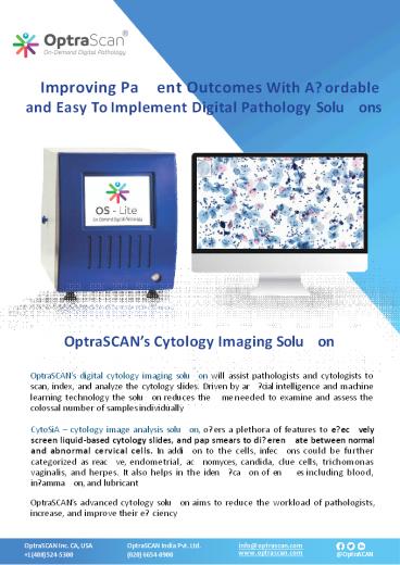
Improving Pa?ent Outcomes With A?ordable and
Easy To Implement Digital Pathology Solu?ons
OptraSCANs Cytology Imaging Solu?on
OptraSCANs digital cytology imaging solu?on will
assist pathologists and cytologists to scan,
index, and analyze the cytology slides. Driven by
ar??cial intelligence and machine learning
technology the solu?on reduces the ?me needed to
examine and assess the colossal number of
samples individually CytoSiA cytology image
analysis solu?on, o?ers a plethora of features to
e?ec?vely screen liquid-based cytology slides,
and pap smears to di?eren?ate between normal and
abnormal cervical cells. In addi?on to the cells,
infec?ons could be further categorized as
reac?ve, endometrial, ac?nomyces, candida, clue
cells, trichomonas vaginalis, and herpes. It
also helps in the iden??ca?on of en??es including
blood, in?amma?on, and lubricant OptraSCANs
advanced cytology solu?on aims to reduce the
workload of pathologists, increase, and improve
their e?ciency
OptraSCAN India Pvt. Ltd. (020) 6654-0900
info_at_optrascan.com www.optrascan.com
2
An ISO 13485 cer??ed company OptraSCAN systems
are CE mark for IVD use All OptraSCAN systems are
for research use only in North America
OptraSCANs Digital Cytology Imaging
Solu?on Extended Depth Of Field OptraSCANs
extended depth of ?eld algorithm generates a
single, en?rely focused composite image to
screen an en?re image e?ec?vely, easily, and
systema?cally. Advantages Rela?vely smaller ?le
size than mul?plane images Quicker to
acquire E?ec?ve and easy naviga?on for accurate
interpreta?on, sharing and archiving Z-Stacking
Technology Stack of images is generated at
di?erent focal planes along the z- axis Viewing
so?ware enables the user to navigate, zoom up and
down the di?erent planes to detect the
three-dimensional regions in focus Can scan
up-to 7 layers Features Of OptraSCANs Volume
Scanning Technology Fully automated mul?layer
scanning at di?erent focal depths. Automated and
User con?gurable focal intervals for Z
stacking Ideal for Rapid On-site evalua?on for
adequacy of cytology sample (Rapid on-site
evalua?on (ROSE)
OptraSCAN India Pvt. Ltd. (020) 6654-0900
info_at_optrascan.com www.optrascan.com
3
An ISO 13485 cer??ed company OptraSCAN systems
are CE mark for IVD use All OptraSCAN systems are
for research use only in North America
OptraSCANs AI ML- based Image Analysis Solu?on
for Cytology Biomarker
Automated computa?on of sample adequacy for the
whole slide cytology image. Iden??ca?on of
abnormal cells and other en??es based on
morphological features and AI based
classi?ca?on. Iden??ca?on of reac?ve,
endometrial, ac?nomyces, candida, clue cells,
trichomonas vaginalis, and herpes
en??es. Iden??ca?on of en??es including blood,
in?amma?on, and lubricant.
Iden??ca?on of Infec?ons Abnormal Cells
Faster iden??ca?on of abnormal cells
OptraSCAN India Pvt. Ltd. (020) 6654-0900
info_at_optrascan.com www.optrascan.com
4
An ISO 13485 cer??ed company OptraSCAN systems
are CE mark for IVD use All OptraSCAN systems are
for research use only in North America
Iden??ca?on of Abnormal Cells
Iden??ca?on of Infec?ons (Ac?nomyces)
Iden??ca?on of HSIL
OptraSCAN India Pvt. Ltd. (020) 6654-0900
info_at_optrascan.com www.optrascan.com
Recommended
CrystalGraphics Presentations


