
Biological Psychology: A Brief History - PowerPoint PPT Presentation
1 / 29
Title:
Biological Psychology: A Brief History
Description:
Biological Psychology: A Brief History puerile, dull, dogmatic, absurd, foolish, and the most frontless piece of charlatanry that the age had produced, while ... – PowerPoint PPT presentation
Number of Views:71
Avg rating:3.0/5.0
Title: Biological Psychology: A Brief History
1
Biological Psychology A Brief History puerile,
dull, dogmatic, absurd, foolish, and the most
frontless piece of charlatanry that the age had
produced, while the "cunning craniologers" who
were beginning to roam the country were seen as
quacks, empirics, manipulating impostors, and
itinerant mountebanks to be looked upon "as
rather knaves than fools." (Cooter, 1984, p. 22)
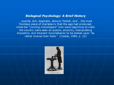
2
Phrenology's Modern Ghosts The quotation above
refers to phrenology, long regarded a
pseudoscientific fraud, but initiated by Franz
Josef Gall, an eminent Viennese physician and
anatomist, who was the first to distinguish the
white and gray matter of the brain. He gathered
masses of data relating "mental and moral"
attributes to the shape of people's heads. He
proposed 26 faculties, or "personality organs,"
each complete and located in specific sites in
the brain. If we have a lot of a trait, such as
facility with language, the brain will be highly
developed in the corresponding area and will
cause the skull to bulge at that point.
3
The phrenologist, also called craniologist,
zoonomist, physiognomist, and other things could
then analyze personality by examining the surface
of the skull. Organ number two is
Philoprogenitiveness, love of children, and is
located at the back of the head, since Gall
believed this area to be prominent in women and
in apes, both of whom supposedly love children
more than do men. Organ number 22 is
Individuality, located immediately above the
nose, since that is what seemed large in the face
of Michelangelo and small in Scottish people!
It may seem that phrenology is ridiculed today,
but if you attend to newspapers and television
you will see that the general point of view
remains popular - we still appear to believe in
mental motives and powers (faculties) that are
localized in specific brain areas. Sometimes
they are conceived as existing in their entirety
in some location - perhaps as "memory units" in
the cerebral cortex. "Didn't someone, Penfield,
I think, revive memories by stimulating the
cortex of patients?" we think. Scientists
Discover Pleasure Centers in the Brain headlined
a Montreal newspaper in the 1950s and variations
on that announcement have appeared frequently in
newspapers and magazines since.
4
You may also have heard of "aggression centers"
in the brain. As recently as the late 1960s and
the 1970s many people were operated upon by
surgeons who believed that there were such
centers (see Valenstein, 1971). We hear also
that there are "feeding and drinking centers" in
the brain and every decade at least two or three
"satiety hormones" are reported, each hoped to be
the chemical that will turn off our hunger and
make us slim. Sometimes it is the "arousal"
center that makes news or dopamine that causes
"awakenings" of Parkinsonism patients. In
countless cases the spirit of phrenology lives on
in the localization of faculties in discrete
brain structures or in the amazing effects of a
new hormone. The same applies to the fruits of
so-called "split brain" research.
5
Richard Gregory noted that laterality
theories, which posit language functions in the
left cerebral hemisphere and spatial apprehension
in the right, tell an old tale One might say
that the current interest in 'cerebral
dominance,' with the left hemisphere of the
cortex supposedly 'analytic' (responsible for
skills such as arithmetic and logical thinking)
and the right hemisphere 'synthetic' or
'analogue' (responsible for intuitive and
artistic skills), is the dying kick of
phrenology. (Gregory, 1987, p. 619-620)
In fact, if we add up all of the claims for
"centers" of one kind or another in the brain, we
could list most of the motives and powers
proposed by the phrenologists. Almost a dozen
separate functions have been attributed to one
brain structure, the hippocampus, and it isn't
even in the cerebral neocortex. Does that
support phrenology?
6
Localization of Function Finding the Right Names
for Functions Let us examine the evidence and
briefly look at the history of research in this
area. We will find that specific parts of the
brain do serve specific functions, as it seems
they must, but it will also be clear that these
functions are often hard to name. That is, when
we list functions like attention, memory,
learning, arousal, hunger, aggression, and the
like, our list seems not to correspond to the
functions built in by the architect of the
nervous system. Happily, recent research
seeking the classic "centers" of the
phrenologists has brought a new understanding of
the characteristics of the nervous system. This
allows a revision of the list of centers, thus
advancing our knowledge of psychology, as well as
of neuroanatomy and physiology.
7
Nineteenth-Century Science The Neural Impulse
Luigi Galvani Alessandro Volta The Rate of
Neural Conduction Hermann Helmholtz
Sensory and Motor Nerves Sir Charles
Bell Francois Magendie Reflex Action Robert
Whytt Marshall Hall Sherrington
Reflexes of the Brain Ivan Sechenov Specific
Nerve Energies Johannes Müller Motor
Cortex Fritsch Hitzig Somatosensory
cortex Bartholomew
8
Merzenich And A New Twist In the
1980s M. M. Merzenich discovered that the
receptive fields in the somatosensory cortex of
the postcentral gyrus are surprisingly malleable.
For example, after amputation of a
finger, the cortical receptive field for that
finger is "invaded" by the receptive fields for
the neighboring digits. This
totally-unexpected malleability of the sensory
cortex the map of the body surface changed
our view of the cortex as a set of fixed maps
influenced by association cortex.
9
Broca's "Language Center" Lesson in
Interpretation The search for brain targets
subserving specific functions appeared to take a
giant leap forward in 1861 when Pierre-Paul Broca
discovered what appeared to be the "speech
center." A patient described as otherwise
normal was unable to articulate language, though
he could understand it. He was called "Tan,"
since "tan-tan" were the only syllables he could
utter. For years he had been paralyzed on the
right side, evidently due to a series of strokes
(to which we refer later). He was referred to
Broca for treatment of an infected bedsore and
soon died. Upon autopsy he was found to have a
lesion in the left hemisphere of the lower
temporal lobe just forward of the temporal lobe
(the inferior posterior frontal lobe) - an area
scarcely four centimeters square. For a century
this area was known as the speech center - its
function turned out to be not what it seemed to
Broca.
10
Reassessing Broca's Finding Karl Pribram
is an eminent neurosurgeon and psychologist who
believes that patients showing "Broca's aphasia"
may suffer from more general disabilities than
the loss of the ability to speak. His
investigation shows that Broca did indeed find
what he was expecting to find. Pribram (1971)
wrote Broca had been taught that language was
a function of the frontal lobes his teachers
derived their doctrine from the phrenologists who
had reasoned that man's high forehead and his
linguistic ability were two of his most
distinguishing features ergo they might well be
related. Broca reasoned that the only place
where his aphasic patient's lesion overlapped the
frontal cortex was in the posterior inferior
portion. Hence Broca's area. Broca's patient
had suffered strokes that involved the middle
cerebral artery this produces widespread damage,
but it was the damage to the frontal cortex,
"Broca's area," that Broca noted. Is the area
the seat of speech?
11
Pribram suggests that it is not Evidence
against Broca's claim is simply that excision of
Broca's area in man's brain, and damage to this
area, has been inflicted without causing any
severe linguistic disturbance... Presumably
therefore all of the lobotomies performed for
psychosurgical reasons (over 10,000) injured
Broca's area to some extent. Yet not a single
report of aphasia due to lobotomy occurred.
(Pribram, 1971) In two catatonic patients who
had not spoken in over twenty years, removal of
Broca's area produced fluent speech that
remained.
12
Is Broca's area the speech center? Pribram went
on to argue that since its removal does not
impair speech and may even improve it, it is not
necessary for speech production. Nonetheless, it
is possible that an intact and malfunctioning
Broca's area may interfere with the motor
production of speech. This, however, is a bit
different from the conventional definition of
"speech center." Penfield's "memories"
suffered a related fate.
13
Penfield's Memories Wilder Penfield
was a Canadian neurosurgeon who operated on a
variety of patients, including epileptics. In
many cases of epilepsy, anti-epileptic drugs are
ineffective and the sufferer is left undergoing
embarrassing and life-threatening seizures, with
no recourse other than a dangerous operation as a
remedy. The operation includes the
removal of portions of cerebral cortex that EEG
analysis has suggested are responsible for the
seizures. In the course of the operation the
patient is awake, while the neurosurgeon probes
for the offending tissue. If signs of a seizure
can be evoked, the cortical area is destroyed
(e.g., by burning it with DC through the
electrode or by aspiration).
14
In the course of these procedures in the
late 1950s, Penfield found that his stimulations,
especially in the parietal and temporal cortex,
often aroused vivid memories in his patients,
complete with color and sound. Often
an incident from many years ago seemed to be
relived, described by the patient as though it
were being played on a videocassette.
Penfield's discovery was described in countless
introductory psychology books and it seemed to
add to the mounting evidence that the
phrenologists, though wrong in detail, were
correct in general. The brain seemed to be
composed of many parts and each part has an
obvious and unique function. Penfield found
stored memories, or so it seemed. We will return
to this issue. What do you think was
going on?
15
Lashley and the Engram In a powerful
display of faith in the precise localization of
function in the brain, a young Karl Lashley
offered to trace the neural connections in the
frog brain and thus determine how the frog brain
worked. He had found some discarded slides of
sections of the frog brain and the neural
connections seemed to him to be traceable. He
was shocked to learn that the stain used was
extremely selective and that the tissue he saw
was therefore a very small fraction of the total.
Despite his discouragement, he spent a
substantial part of the rest of his life trying
to accomplish that mission. His findings had a
profound and lasting influence on the search for
brain-behavior relations Thompson Robinson,
1978). In 1929 he published a monograph, Brain
Mechanisms and Intelligence, detailing his
findings concerning the effects of brain lesions
on the ability of rats to learn mazes. What did
he find?
16
What he essentially found was that quite
a bit of the cortex had to be destroyed before
any deficit was found and, surprisingly, that it
did not matter from what part of the cortex
tissue was destroyed. Thus, such and such a
deficit would be found on the most difficult maze
with destruction of 50 of the cortex. But it
did not seem to matter what 50 was destroyed, as
long as the primary projection areas were spared.
A rat that had one large lesion comprising half
its cortex would perform similarly to a rat that
had twenty small lesions scattered over the
cortex. What was important was only the
percentage destroyed, not its location. This
means that the cortex is equipotential any part
can carry out the function of any other part,
within limits. The degree of the deficit did
increase with the size of the lesion, however,
which led Lashley to propose the principle of
mass action. This means that cortical tissue may
be equipotential, but that its efficiency depends
on the amount which is remaining - the mass of
cortex available is important.
17
The 1929 report was very discouraging to
researchers aiming to show localization of
function. in the brain. If the particular locus
of brain damage is not crucial, then how can
specific memories be stored in specific places?
Whether the memory trace (or engram) consists of
neural circuits, concentrations of proteins such
as RNA, or presence of neurotransmitters, the
location of the lesions should be very
important. Lashley continued his
search for a great many years, a search that he
described in 1950 ("In Search of the Engram").
After his 1929 monograph he tried slicing the
cortices of his subjects, so that their brains
resembled sliced hams, only to find no deficits
in learning tasks. He destroyed the linkage
between the sensory and the motor areas and even
lesioned the cerebellum. The cerebellum
influences motor behavior and, since other
lesions had little effect, maybe the engrams
required to learn mazes were stored their. But
even those subjects, whose movements were
hampered and who crawled, rolled, and squirmed
along the alley, came to the choice points and
rolled down the correct alley. How can that be?
18
Lashley's Legacy Other data corroborate
Lashley's basic findings and two authoritative
reviewers (Pribram, 1971 Thompson Robinson,
1977) agree that his findings were legitimate.
Instances of serious brain damage producing
little deterioration in performance abound.
For example, Chow (1970) destroyed
three-quarters of the visual cortex of cats and
at the same time cut more than three-quarters
through their optic nerves. Such an operation
would reduce the animal's vision to near nil, one
would think, and it does cause disruption of a
previously-learned visual discrimination. But
the cats relearned the discrimination as quickly
as they had originally learned! Other data show
the discrimination performance of cats and other
animals to be passable after even greater
destruction of the visual pathway. It seems that
a few hundred visual cortical cells is sufficient
for the learning of fairly difficult visual
discriminations (Lashley, 1970).
19
Is Your Brain Really Necessary? That
was the title of a piece appearing in Science
(Lewin, 1980). It describes the findings of a
British neurologist, John Lorber, at the
University of Sheffield in the United Kingdom.
Lorber's research involves hydrocephalics, whose
brain ventricles accumulate an excess of
cerebrospinal fluid. When this occurs
in an infant or young child, the skull expands to
make room for the excess fluid. But in older
children and adults, the skull is not malleable
and the fluid crushes the forebrain against the
inside of the skull. In many instances great
brain damage occurs, accompanied by grave
disturbances in function. However, in a great
many cases, there is no obvious deficit, even
though the brain damage is extreme. As Lorber
put it
20
There's a young student at this university, who
has an IQ of 126, has gained a first-class honors
degree in mathematics, and is socially completely
normal. And yet the boy has virtually no
brain...When we did a brain scan on him, we saw
that instead of the normal 4.5-centimeter
thickness of brain tissue between the ventricles
and the cortical surface, there was just a thin
layer of mantle measuring a millimeter or so.
His cranium is filled mainly with cerebrospinal
fluid. (Lewin, 1980, p. 1232)
The eminent British neuroanatomist ,
Patrick Wall, at University College, London,
commented that "Scores of similar accounts litter
the medical literature, and they go back a long
way." He praised Lorber for compiling a
remarkable set of data, rather than relying on
mere anecdotal accounts. Wall wondered how we
may account for such findings. How
indeed may we account for them? If Lorber is
right and if Lashley's search means anything,
then we must at least question the old
supposition that the cerebral cortex is the seat
of all intelligent behavior and particularly that
it is the repository for precisely localized
memories. But what of Penfield's famous
findings?
21
Penfield Reconsidered Penfield was no doubt
sincere in his belief that he had found the
anatomical substrate for memory, but Valenstein
(1973) showed that more recent evidence paints a
somewhat different picture. Fedio and Van Buren
(1971), at what was then the National Institute
for the Study of Neurological Disease and Stroke,
point out that many surgeons use precisely the
procedure used by Penfield, yet no one seems to
have found revived memories as he reported. That
fact, along with the report by Mahl (1970) that
such patients often do report seeing flashes of
light or hearing brief sounds, suggests another
interpretation for Penfield's finding.
Imagine yourself as a patient under the
conditions experienced by his patients. You are
undergoing a brain operation and, while you were
under anesthesia, the surgeon has cut through the
scalp, the skull, and the dura under the skull.
You are now sitting there awake and the surgeon
is touching your brain with a stimulating
electrode! It is surely fair to say that you
might be a little "on edge" or "reactive" under
such circumstances.
22
Suppose now that during the stimulation you see
a flash of light or hear a sound, just as you
might see flashes and hear "bells ring" when
something strikes you in the head. You say, "I
heard something," and the surgeon asks what it
was. Was it like a train whistle? Yes, it was
and you add, "I can see the station and there is
my mother..." and so on. With a little prodding,
completely inadvertently done by the surgeon, a
patient may well tell many stories under such
circumstances. Does this amount to the revival
of memories? In a sense it does, but the
stimulation of the brain surface appears to
arouse only light flashes and brief auditory
sensations. The elaborations of these are
aroused by the questions of the surgeon in the
context of a highly reactive subject. Penfield
was entirely well meaning and no reader of his
biography, No Man Alone (1970) could believe that
he was intentionally perpetuating a fraud. He
found what his education had led him to expect
and one cannot blame him for believing that he
found it. But others did not find it and the
reason for their failure is clear.
23
Motivation and Emotion Thou shalt not sit With
statisticians nor commit A social
science (Auden, 1946, st. 27) Probably
the best known discoveries in physiological
psychology occurred in the area of motivation and
emotion this includes the discovery of what some
called "reward" and "punishment" centers, feeding
centers, aggression centers, and the like. The
story involves the limbic system, particularly
the hypothalamus. DRAW DIAGRAM
If the brain were an apple, its core would
be the limbic system, old cortex arranged
essentially the same in us as it is in dogs,
rabbits, and rats. Limbic means "border" and
refers to the brain tissue bordering the midline
of the brain. The hypothalamus, a cluster of
cell bodies about as large as the tip of your
thumb, is a crucial part of this system and
controls the autonomic nervous system.
24
The autonomic nervous system is subdivided into
the sympathetic and the parasympathetic branches.
The division was suggested by two Viennese
neurologists, Karplus and Kreidl, in 1909 and
subsequent research has supported their view.
They suggested that the anterior (forward)
portion of the hypothalamus controls
parasympathetic activity this includes
conservative functions such as sleep, sexual
activity, feeding, and other "vegetative"
functions. On the other hand, the posterior
(rearward) hypothalamus controls aggression,
flight, and other activities that use up energy
in the interests of survival. The posterior
hypothalamus increases sympathetic activity,
which means that heart rate increases, blood is
shunted from the viscera to the muscles, the
lungs exchange gases more rapidly, and the
affected organism is more able to fight for its
life or to flee. And Karplus and Kreidl were
correct, as many studies since have shown. The
hypothalamus seems to be a center for motivation
and emotion and in a sense it is.
25
Papez's Circuit In 1936 a
neuroanatomist, James Papez, made a bizarre
proposal, based wholly on anatomical evidence and
reasonable assumptions about the nervous system
and emotion. EXPLAIN We now know
that stimulation of the brainstem reticular
formation (Moruzzi Magoun, 1949 Hebb, 1954)
produces nonspecific arousal of the entire
forebrain. EXPLAIN In 1942
Hetherington and Ranson found what came to be
called the "satiety center. EXPLAIN
26
In 1951 Anand and Brobeck at Yale
University found the "feeding center." While
attempting to insert stimulating electrodes into
the amygdala of rats (the amygdala is covered
below), they inadvertently destroyed the lateral
nuclei of the hypothalamus and found that their
subjects died after the operation. The cause of
death was aphagia the rats refused to eat and
spat out food that was forced in their mouths.
Thus, the hypothalamus seemed to control both
eating and not eating. If the VM "satiety"
center were damaged, hyperphagia, or gross
overeating, occurred. If the lateral nuclei (LH)
were destroyed, eating ceased. Reasonably
enough, when the VM was stimulated, eating
ceased, and stimulation of the LH produced
eating. Here were the feeding and satiety
centers and everyone accepted them. The only
question was what new centers would be discovered
to account for new behaviors? No wonder Hess won
the Nobel Prize.
27
Re-evaluation of LH and VMH Function
A great deal of evidence indicates that the
effects of VM and LH lesions are more general
than had been believed and that the ordinary
functions of the two structures are inhibitory
and excitatory, respectively. Destruction of the
VM removes an inhibitory influence and leads to
increased responsiveness to strong external cues.
The LH appears excitatory, so that
stimulation of it appears in some ways to act as
does destruction of the VM. Such stimulation can
maintain lever pressing, produce feeding, cause
stalking of prey, and other activities that seem
describable as "outer directed." Destruction of
the LH causes decreased responsiveness to
external stimuli and even to one's own body.
Operated cats will remain immobile, even when
placed in uncomfortable positions, and will
ignore stimuli that would ordinarily elicit
strong reactions. This sensory neglect
is a powerful general effect, yet, as Carlson
(1991) noted, the lateral hypothalamus was called
the "feeding center" for approximately two
decades! Therein lies a lesson for us.
28
Add to This List (see MIT Press History Chapter
10 online at the Nuoro page of www.geocities.com/m
alonejc2007. There is far more detail than will
appear in the actual book.) The Amygdala as
aggression center Kluver/Bucy Effect and
psychosurgery Effects of bilateral
amygdalectomy Freeman Watts
Downers finding James Olds and pleasure
centers Hebb Delgado Valenstein
29
- Brocas Language Center (motor control)
- Penfields Memories (surgeons
prompting) - Morruzi Magouns Arousal Center (EEG
only) - Satiety and Feeding Centers (E/I control)
- Aggression Centers (psychic blindness)
- Pleasure/Reward Centers (also E/I
control) - In all of these cases, misinterpretations
of effects were made and, much later, a
better-informed interpretation arose. - So..beware of claims of breakthroughs!
- In all of these cases,
Recommended
CrystalGraphics Presentations


