
Anatomy - PowerPoint PPT Presentation
1 / 67
Title: Anatomy
1
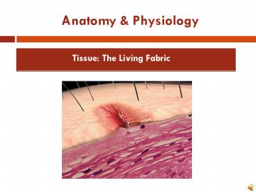
Anatomy Physiology
- Tissue The Living Fabric
2
Anatomy Physiology
- Tissue The Living Fabric
- Objectives
- Identify and describe the functions of the 4 main
types of body tissues - Describe the various types and functions of
epithelia - Explain the properties and functions of different
types of connective tissue - Identify the major types of muscle tissue
- Describe the basic types and functions of nerve
tissue
3
Tissues
- Groups of cells similar in structure and function
- Types of tissues
- Epithelial tissue
- Connective tissue
- Muscle tissue
- Nerve tissue
4
Nervous tissue Internal communication Brain,
spinal cord, and nerves
Muscle tissue Contracts to cause movement
Muscles attached to bones (skeletal) Muscles of
heart (cardiac) Muscles of walls of hollow
organs (smooth)
Epithelial tissue Forms boundaries between
different environments, protects, secretes,
absorbs, filters Skin surface (epidermis)
Lining of GI tract organs and other hollow organs
Connective tissue Supports, protects,
binds other tissues together Bones Tendons
Fat and other soft padding tissue
Intro to tissues
Figure 4.1
5
Epithelial Tissue (Epithelium)
- Two main types (by location)
- Covering and lining epithelia
- On external and internal surfaces
- Glandular epithelia
- Secretory tissue in glands
6
Functions of Epithelial Tissue
- 6 main functions of epithelium
- Protection (skin)
- Absorption (digestive tract, kidneys)
- Filtration (digestive tract, kidneys)
- Excretion (digestive tract, kidneys)
- Secretion (glands, kidneys)
- Sensory reception (skin)
7
Homeostatic Imbalance
- Epithelial Tissue
- An important characteristic of cancerous
epithelial cells is their failure to respect the
basement membrane boundary. (85 out of 100
cancers are of epithelial cells) - They penetrate it and invade the tissues beneath.
8
Characteristics of Epithelial Tissue
- Cells have polarity- apical (upper, free) and
basal (lower, attached) surfaces- cell regions
near the apical surface differ in structure and
function from cell regions in the basal surface. - Apical surfaces may have
- Microvilli finger-like extensions of the plasma
membrane that increase surface area. - Lining of the intestine
- Cilia tiny hair like projections that propel
substances along their free surface. - Lining of the trachea
9
Characteristics of Epithelial Tissue
- Are composed of closely packed cells to form
continuous sheets. - Supported by a connective tissue reticular lamina
(under the basal lamina) - A layer of extracellular material containing a
fine network of collagen and protein fibers. - Avascular (contains no blood vessels) but
innervated (supplied by nerve fibers). - Epithelial cells are nourished by substances
diffusing from blood vessels in the underlying
connective tissue. - High rate of regeneration
- Reproduce rapidly to replace lost cells due to
hostile substances in the external environment.
10
Classification of Epithelia
- Ask two questions
- How many layers?
- 1 layer
- simple epithelium
- Very thin most concerned with absorption,
secretion, and filtration. - 2 or more layers stratified epithelium
- More durable, major role is protection.
Figure 4.2a
11
Classification of Epithelia
- What type of cell?
- Squamous
- Flattened and scale-like
- Cuboidal
- Box-like, tall as they are wide
- Columnar
- Tall and column shaped
- (If stratified, name according to apical (top)
layer of cells)
Figure 4.2b
12
Epithelia Simple Squamous
- Thin, often permeable cells
- Found where filtration and exchange of substances
by rapid diffusion is a priority - Kidneys
- Lungs
- Blood vessels
13
Epithelia Simple Squamous
- Two noteworthy names
- Endothelium innercovering slick-friction
reducing lining - The lining of lymphatic vessels, blood vessels,
and heart - Mesothelium middlecovering
- The epithelium of serous membranes in the ventral
body cavity
14
Figure 4.3a
15
Figure 4.3b
16
Figure 4.3c
17
Figure 4.3d
18
Figure 4.3e
19
Epithelia Stratified Cuboidal
- Quite rare in body
- Found in some sweat and mammary glands
- Typically two cell layers thick
Sweat Duct
20
Epithelia Stratified Columnar
- Limited distribution in body
- Small amounts in pharynx, male urethra, and
lining some glandular ducts - Also occurs at transition areas between two other
types of epithelia
21
(f) Transitional epithelium
Description Resembles both stratified squamous
and stratified cuboidal basal cells cuboidal or
columnar surface cells dome shaped or
squamouslike, depending on degree of organ
stretch.
Transitional epithelium
Function Stretches readily and permits
distension of urinary organ by contained urine.
Location Lines the ureters, urinary bladder,
and part of the urethra.
Basement membrane
Connective tissue
Photomicrograph Transitional epithelium lining
the urinary bladder, relaxed state (360X) note
the bulbous, or rounded, appearance of the cells
at the surface these cells flatten and become
elongated when the bladder is filled with urine.
Figure 4.3f
22
Answer the following questions and turn in!! ?
- Explain what is meant by epithelial tissue being
avascular but innervated.
- What structure can be noted on the apical surface
of the cells in this image? - What is the name of this tissue type?
- A multilayered epithelium with cuboidal basal
cells and flat cells at its surface would be
classified as ________?
23
Answer the following questions and turn in!! ?
- Explain what is meant by epithelial cells having
polarity
- What is significant about the cells closes to the
basement membrane of this tissue type? - What is the name of this tissue type?
- A multilayered epithelium with cuboidal basal
cells and columnar cells at its surface would be
classified as ________? - What types of organs would you find transitional
epithelium in?
24
Glandular Epithelia
- Objectives
- Define gland
- Differentiate between exocrine and endocrine
glands, and differentiate between multicellular
and unicellular glands - Describe how multicellular exocrine glands are
classified structurally and functionally
25
Glandular Epithelia
- A gland is one or more cells that makes and
secretes an aqueous fluid. - Glandular cells obtain needed substances from
blood and transform them chemically into a
product that is then released from the cell. - Classified by
- Site of product releaseendocrine (internally
secreting) or exocrine (externally secreting) - Relative number of cells forming the
glandunicellular (e.g., goblet cells) or
multicellular (typically ducted)
26
Endocrine Glands
- Ductless glands
- Secrete hormones that travel through lymph or
blood to target organs
27
Exocrine Glands
- More numerous than endocrine glands
- Secrete products into ducts
- Secretions released onto body surfaces (skin) or
into body cavities - Examples include mucous, sweat, oil, and salivary
glands
28
Unicellular Exocrine Glands
Goblet Cell
- The only important unicellular gland are mucous
cells and goblet cells. (scattered) - Goblet cells- look like a glass with a stem due
to the accumulation of mucin at the top of the
cell. - Both produce mucin that dissolves in water when
secreted and forms mucous- a slimy protective and
lubricating coating found within the human body.
29
Multicellular Exocrine Glands
- Multicellular exocrine glands are composed of a
duct and a secretory unit - Classified according to
- Duct type
- Simple unbranched duct
- Compound branched duct
- Structure of their secretory units
- Tubular secretory cells form tubes.
- Alveolar (acinar) secretory cells form small
sacs. - Tubuloalveolar have both types of secretory
units.
30
Figure 4.5
31
Modes of Secretion
- Merocrine
- Products are secreted by exocytosis
- Examples pancreas, sweat and salivary glands.
- Holocrine
- Products are secreted by rupture of gland cells
- Example sebaceous glands
32
Answer the following questions and turn in!! ?
- What is a gland?
- Explain the difference between endocrine and
exocrine glands and provide an example of each - What type of an exocrine gland is a goblet cell
and what does it produce? - Draw and label the various structural
presentations for multicellular exocrine glands. - What is the primary difference between the way
merocrine and halocrine glands secrete their
products?
33
Connective Tissue
- Objectives
- Identify common characteristics of connective
tissue, and list and describe its common
structural elements - Describe the common types of connective tissue
found in the body and indicated their particular
functions
34
Connective Tissue
- Most abundant and widely distributed tissue type
- Four classes
- Connective tissue proper
- Cartilage
- Bone tissue
- Blood
35
(No Transcript)
36
Major Functions of Connective Tissue
- Binding and support
- Protection
- Insulation
- Transportation (blood)
37
Characteristics of Connective Tissue
- Connective tissues have
- Mesenchyme (embryonic tissue) as their common
tissue of origin - Varying degrees of vascularity (supply of blood
vessels) - Cells separated by nonliving extracellular matrix
(ground substance and fibers) - This is what enables the connective tissue to
bear weight, withstand great tension, and endure
physical trauma.
Mesenchymal Cells
Areolar Connective Tissue
38
Structural Elements of Connective Tissue
- Ground substance
- Unstructured material that fills the space
between the cells and contains the fibers - Functions as a molecular sieve through which
nutrients and other dissolved substances diffuse
between blood capillaries and cells. - Components
- Interstitial fluid
- Adhesion proteins (glue)
- Proteoglycans
- Protein core large polysaccharides
(chrondroitin sulfate and hyaluronic acid) - Trap water in varying amounts, affecting the
thickness of the ground substance
39
Structural Elements of Connective Tissue
- Fibers provide support.
- Three types of fibers
- Collagen (white fibers)
- Strongest and most abundant type
- Provides high tensile strength
- Elastic
- Networks of long, thin, elastin fibers that allow
for stretch - Reticular
- Short, fine, highly branched collagenous fibers
40
Structural Elements of Connective Tissue
- Cells
- Mitotically active and secretory cells blasts
- Mature cells cytes
- Fibroblasts in connective tissue proper
- Chondroblasts and chondrocytes in cartilage
- Osteoblasts and osteocytes in bone
- Hematopoietic stem cells in bone marrow
- Fat cells, white blood cells, mast cells, and
macrophages
41
(No Transcript)
42
Connective Tissue Embryonic
- Mesenchymeembryonic connective tissue
- Gives rise to all other connective tissues
- Gel-like ground substance with fibers and
star-shaped mesenchymal cells
43
Answer the following questions and turn in!! ?
- What are the 4 classes of connective tissue?
- What 3 types of fibers provide support for
connective tissue? - What are 4 functions of connective tissue?
- What function(s) does adipose serve?
44
Connective Tissue Proper
The name connective tissue proper is used to
designate the connective tissue that fills
interstitial spaces as opposed to the specialized
connective tissues (blood, bones, cartilage,
etc).
- Types
- Loose connective tissue
- Areolar
- Adipose
- Reticular
- Dense connective tissue
- Dense regular
- Dense irregular
- Elastic
45
Figure 4.8a
46
Figure 4.8b
47
Figure 4.8c
48
Figure 4.8d
49
Figure 4.8e
50
Figure 4.8f
51
Connective Tissue Cartilage
- Three types of cartilage
- Hyaline cartilage
- Elastic cartilage
- Fibrocartilage
52
Homeostatic Imbalance
- Cartilage
- Aging cartilage cells lose their ability to
divide ? injure cartilages heal slowly. - During later life cartilages tend to calcify or
ossify (become bony).
53
Figure 4.8g
54
Figure 4.8h
55
Figure 4.8i
56
Other tissues
57
Figure 4.8j
58
(k) Others blood
Description Red and white blood cells in a fluid
matrix (plasma).
Plasma
Function Transport of respiratory gases,
nutrients, wastes, and other substances.
Neutrophil
Location Contained within blood vessels.
Red blood cells
Lymphocyte
Photomicrograph Smear of human blood (1860x)
two white blood cells (neutrophil in upper left
and lymphocyte in lower right) are seen
surrounded by red blood cells.
Figure 4.8k
59
Nervous Tissue
- Nervous system
Figure 4.9
60
Muscle Tissue
Figure 4.10a
61
Muscle Tissue
Figure 4.10b
62
Muscle Tissue
Figure 4.10c
63
Epithelial Membranes
- Cutaneous membrane (skin)
- Consists of keratinized stratified squamous
epithelium (epidermis) firmly attached to a thick
layer of dense irregular connective tissue
(dermis)
64
Epithelial Membranes
- Mucous membranes
- Mucosae
- Line body cavities open to the exterior (e.g.,
digestive and respiratory tracts)
65
Epithelial Membranes
- Serous Membranes
- Serosaemembranes (mesothelium areolar tissue)
in a closed ventral body cavity - Parietal serosae line internal body walls
- Visceral serosae cover internal organs
66
Steps in Tissue Repair
67
Steps in Tissue Repair
68
Steps in Tissue Repair
69
Homeostatic Imbalance
- Scar Tissue
- Scar tissue that forms in any muscular organ-
heart or urinary bladder can severely impair the
function of that organ. - Scars reduce the internal volume of the organ and
block movement of substances through the hollow
organ. - Can hamper the muscles ability to contract.
- Heart progressive heart failure.
- Visceral organs adhesions connect organs
together and prevent normal shifting about ? ex.
Intestines adhesions obstruct the flow of
foodstuffs. - Tissue Damage-Snake Bite
70
The End! ?
71
Developmental Aspects
- Primary germ layers ectoderm, mesoderm, and
endoderm - Formed early in embryonic development
- Specialize to form the four primary tissues
- Nerve tissue arises from ectoderm
- Muscle and connective tissues arise from mesoderm
- Epithelial tissues arise from all three germ
layers
Recommended
CrystalGraphics Presentations


