
The Axial Skeletal System - PowerPoint PPT Presentation
1 / 117
Title:
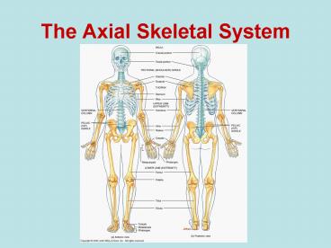
The Axial Skeletal System
Description:
Title: Slide 1 Author: Skish Last modified by: Lori McLoughlin Created Date: 10/5/2006 1:49:06 AM Document presentation format: On-screen Show (4:3) – PowerPoint PPT presentation
Number of Views:116
Avg rating:3.0/5.0
Title: The Axial Skeletal System
1
The Axial Skeletal System
2
Divisions of the Skeletal System
- Humans are born with approximately 300 bones
which fuse to 206 bones as adults. - There are 2 main divisions of the skeletal
system axial skeleton and appendicular skeleton.
3
Divisions of the Skeletal System
- Axial skeleton forms the vertical axis of the
body. - 80 bones skull (22), vertebral column (26),
ribcage (25), auditory ossicles (6), and hyoid
(1) - Appendicular skeleton forms the arms, legs and
the girdles - Girdles attach the arms and legs to the axial
skeleton - 126 bones pectoral girdle (4), arms (60), legs
(60) and pelvic girdle (2)
4
(No Transcript)
5
Skull
- Superior end of the vertebral column
- Composed of flat and irregular shaped bones
- Large hollow space within the skull is called the
cranial vault or cranial cavity. - Functions to
- Surround and protect the brain
- Be points of attachment for the facial muscles
(landmarks)
6
Divisions of the Skull
- Cranial division consists of 8 flat bones that
form a protective box around the brain. - Help to form the cranial vault (cavity)
- Frontal (1) forms the anterior portion of the
cranial cavity - Forms the superior orbits of the eyes and forms
the forehead
7
Frontal Bone Landmarks
- Supraorbital margin a thickened ridge of bone
found superior to the orbit of the eye. - Just deep to the eyebrow and more prominent on
the lateral portion - Point for muscle attachment (PFMA)
- Supraorbital foramen a small opening found on
the medial aspect of the supraorbital margin. - Can feel it best inferior to the margin
- Allows blood vessels and nerves to enter the
frontal bone
8
Frontal Bone Landmarks
- Frontal sinus A hollow space found within the
frontal bone, superior and medial to the
supraorbital margin. - Can only be seen with a sagittal cut
- ¼ inch superior to the eyebrows
- House mucus and macrophages for trapping and
destroying foreign particles.
9
Frontal Bone Landmarks
- Frontal sinus A hollow space found within the
frontal bone, superior and medial to the
supraorbital margin. - Can only be seen with a sagittal cut
- ¼ inch superior to the eyebrows
- House mucus and macrophages for trapping and
destroying foreign particles.
10
(No Transcript)
11
(No Transcript)
12
(No Transcript)
13
(No Transcript)
14
(No Transcript)
15
(No Transcript)
16
(No Transcript)
17
(No Transcript)
18
(No Transcript)
19
(No Transcript)
20
(No Transcript)
21
(No Transcript)
22
Parietal Bones (2)
- Form the lateral walls and the superior portion
of the cranium. - Landmarks
- Temporal fossa A large, shallow depression that
begins on the parietal bone and extends to the
frontal bone. - PFMA
23
Temporal Bones (2)
- Form the inferior lateral walls and a portion of
the floor of the cranium. - Articulate with the mandible (lower jaw) to form
the temporomandibular joint (TMJ) - Temporal bone landmarks
- Mastoid process A large, blunt projection found
posterior to the external auditory meatus. - Bump behind the ear
- PFMA
24
Temporal Bone Landmarks cont..
- Styloid process A thin, sharp projection found
inferior and medial to the external auditory
meatus. - Covered with muscle so it is more difficult to
identify - PFMA
- Zygomatic process A thin, flat projection found
anterior to the external auditory meatus. - PFMA
- External auditory meatus The external ear canal
- Opening through which the auditory nerve runs.
25
Temporal Bone Landmarks cont..
- Mandibular fossa A shallow depression found
inferior and slightly anterior to the external
auditory meatus - This forms an articulation with the mandible
- Easy to see inferiorly if the mandible is removed
26
Occipital Bone (1)
- Forms the posterior wall and the floor of the
cranium. - The spinal cord passes through this as it exits
the cranial vault. - Occipital bone landmarks
- External occipital protuberance A prominent
midline projection found on the superior surface. - Where the occipital bone turns to form the
horizontal part.
27
Occipital Bone Landmarks cont..
- Superior nuchal line Two curved ridges that
extend laterally from the external occipital
protuberance. - PFMA
- Inferior nuchal line Two curved ridges that
extend laterally from the external occipital
protuberance, inferior to the superior nuchal
line. - PFMA
- Foramen magnum A large opening in the inferior
surface of the occipital bone that allows the
spinal cord to exit the cranial cavity. - Largest foramen in the body.
28
Occipital Bone Landmarks cont..
- Occipital condyles Paired oval-shaped
projections found lateral to the foramen magnum. - Form an articulation with the 1st bone of the
spine (atlas)
29
(No Transcript)
30
Pause and Practice!Number 1-10
31
2
1
32
4
5
3
33
7
6
34
8
9
10
35
Sphenoid Bone (1)
- Forms the anterior floor of the cranial cavity.
- Also forms a portion of the lateral walls of the
cranial cavity. - Forms the posterior wall of the orbits of the
eyes. - The keystone bone for the cranium because it
articulates with all other cranial bones. - Shape resembles a bat with outstretched wings
when viewed superiorly.
36
Sphenoid Bone Landmarks
- Greater wing The larger, inferior projection of
the sphenoid that forms a portion of the floor
and the lateral walls of the cranium. - Also forms the posterior wall of the orbits of
the eyes. - Lesser wing The smaller, superior projection of
the sphenoid bone located posterior to the
frontal bone.
37
Sphenoid Bone Landmarks cont..
- Sella turcica A small, saddle-like depression
found between the greater and lesser wings that
surrounds and protects the pituitary gland. - Pituitary is an important endocrine gland
- 3 parts to the sella turcica
- Tuberculum sellae The anterior portion of the
sella turcica. - Closest to the lesser wing.
38
Sphenoid Bone Landmarks cont..
- 3 parts to the sella turcica continued
- Hypophyseal fossa The seat of the saddle.
- Where the pituitary gland resides
- Dorsum sellae The posterior portion of the sella
turcica. - Closer to the greater wing
39
(No Transcript)
40
(No Transcript)
41
(No Transcript)
42
(No Transcript)
43
(No Transcript)
44
Ethmoid Bone (1)
- The small bone located anterior to the sphenoid
bone in the middle of the frontal bone. - Forms a small portion of the anterior floor of
the cranium. - Also forms a small portion of the medial wall of
the eye orbits. - Also forms the superior portion of the nasal
septum.
45
Ethmoid Bone Landmarks
- Cribriform plate Paired projections found
lateral to the crista galli. - Has small openings called the olfactory foramina.
- Olfactory foramina A series of small openings
found within the cribriform plate that allow
nerves from the olfactory epithelium to pass
directly into the brain. - These nerves give us our sense of smell.
46
Ethmoid Bone Landmarks cont..
- Crista galli A small triangular projection found
in the center of the ethmoid bone. - Near the front of the cranial cavity.
- Point of attachment for the meninges (protective
coverings of the brain). - Perpendicular plate A small vertical projection
arising from the inferior surface of the ethmoid
bone. - Forms the superior portion of the nasal septum.
- Articulates with the vomer (facial bone).
47
Ethmoid Bone Landmarks cont..
- Superior and middle nasal conchae Two thin,
scroll-shaped projections found lateral to the
perpendicular plate - The middle nasal conchae is inferior to the
superior nasal conchae. - These increase surface area of the nasal
passageways - Ethmoidal cells Air spaces found within the
lateral masses of the ethmoid bone. - Small sinuses
48
(No Transcript)
49
(No Transcript)
50
(No Transcript)
51
(No Transcript)
52
(No Transcript)
53
Sutures
- Fibrous joints found between the bones of the
cranium. - There are 4 major sutures
- Coronal unites the frontal bone and both
parietal bones - Sagittal unites the two parietal bones on the
superior midline of the skull - Lambdoid unites the two parietal bones to the
occipital bone. - Squamous (2) unite the parietal and temporal
bones on the lateral sides of the skull
54
Pause and Practice Again!Number 1-10
55
1
2
56
3
4
57
5
6
58
7
8
59
9
10
60
(No Transcript)
61
(No Transcript)
62
(No Transcript)
63
The Facial Division
- A group of 14 irregular bones that serves as
points of attachment for muscles of the face. - Nasal (2) form the bridge of the nose.
- Rectangular shaped bones
- PFMA
- Maxillae (2) Form the upper jaw.
- Articulate with every face bone except the lower
jaw. - Form part of the floors of the orbits, lateral
walls and floor of the nasal cavity, and most of
the hard palate (bony roof of the mouth).
64
Maxillary Landmarks
- Infraorbital foramen Small openings found
inferior to the orbits of the eyes. - Allows passage of blood vessels and nerves.
- Palatine process a lateral projection that forms
one half of the anterior portion of the hard
palate. - Typically the 2 processes unite during weeks
10-12 of embryo development. If not, cleft
palate will result. This negatively impacts
speech and swallowing.
65
Maxillary Landmarks cont
- Maxillary sinuses a series of small spaces
within the maxillae. - Empty into the nasal cavity.
66
(No Transcript)
67
(No Transcript)
68
(No Transcript)
69
Zygomatic Bone (2)
- Form the prominence of the cheeks.
- Also form part of the lateral wall and floor of
each orbit. - Articulate with the frontal, maxilla, sphenoid
and temporal bones. - Zygomatic bone landmarks
- Temporal process a thin, flat projection arising
from the lateral, posterior surface of the
zygomatic bone. - Articulates with the zygomatic process of the
temporal bone.
70
Zygomatic Bone Landmarks cont..
- Zygomatic arch created by the articulation of
the temporal process of the zygomatic bone and
the zygomatic process of the temporal bone.
71
(No Transcript)
72
Lacrimal Bones (2)
- The smallest bones of the facial division.
- Resemble the shape and size of a fingernail
- Posterior and lateral to the nasal bones and form
part of the medial wall of each orbit. - Lacrimal bone landmark
- Lacrimal fossa a small vertical groove formed
with the maxilla, that helps drain fluid away
from the eye. - Houses a lacrimal sac that gathers tears and
passes them into the nasal cavity.
73
(No Transcript)
74
(No Transcript)
75
- Palatine Bones (2) L shaped bones that form
the posterior portion of the hard palate. - The parts that make-up the hard palate are called
horizontal plates. - Inferior Nasal Conchae (2) scroll shaped bones
that form a portion of the inferior, lateral
walls of the nasal cavity. - Increase surface area and help filter air along
with the superior and middle nasal conchae of the
ethmoid bone.
76
(No Transcript)
77
(No Transcript)
78
(No Transcript)
79
(No Transcript)
80
- Vomer (1) a triangular bone that forms a portion
of the posterior floor of the nasal cavity. - Articulates with the perpendicular plate of the
ethmoid bone to form the inferior portion of the
bony nasal septum. - Mandible (1) the largest bone of the facial
division. - Except for the ossicles, it is the only moveable
skull bone.
81
Mandibular Landmarks
- Mandibular body a triangular bone that forms a
portion of the posterior floor of the nasal
cavity. - Ramus the short, vertical portion of the
mandible. - Angle the area where the ramus and the body of
the mandible meet. - Coronoid process a small triangular projection
found on the superior anterior portion of the
ramus.
82
Mandibular Landmarks cont
- Condylar process a small rounded projection
found on the superior posterior portion of the
ramus. - Articulates with the mandibular fossa to create
the temporomandibular joint (TMJ). - Mental foramen small openings found in the
anterior surface of the body of the mandible that
allow blood vessels and nerves to enter the
mandible. - Alveoli sockets for teeth
83
(No Transcript)
84
Auditory Ossicles
- The 6 smallest bones in the human body.
- Located medial to the eardrum.
- Connected by synovial joints.
- Function to transfer sound waves from the eardrum
to the inner ear. - The bones are as follows
- Malleus- attaches to the eardrum and is commonly
called the hammer. - Incus- middle bone that is commonly called the
anvil. - Stapes- Smallest bone and is commonly called the
stirrup.
85
(No Transcript)
86
(No Transcript)
87
Hyoid Bone
- Located superior to the larynx (voice box).
- U shaped
- The only bone that does not articulate with
another bone. - Suspends from the styloid processes by ligaments
and muscles. - Often fractured during strangulation.
- Functions to support the tongue.
88
(No Transcript)
89
(No Transcript)
90
The Vertebral Column
- Also called the spine, backbone or spinal column.
- Consists of 33 (children) or 26 (adults) bones
called vertebrae. - Functions to protect the spinal cord, support the
head, and serve as attachment points for the
ribs, pelvis, back muscles and arm muscles.
91
Vertebrae
- Vary in size, shape and detail but have many
similarities. - Consist of 3 main parts vertebral body,
vertebral arch and several processes. - Vertebral body the thickened anterior portion of
a vertebra. - Holds the intervertebral disc and contains
foramina for the entrance of blood vessels. - Intervertebral discs pads of fibrocartilage that
help hold the vertebrae in place. - Compress throughout the day due to weight and
water loss. This compression does not change
height as we age.
92
(No Transcript)
93
Vertebrae continued
- Vertebral arch located posterior to the
vertebral body. - Forms the vertebral foramen with the vertebral
body. - The vertebral arch consists of the pedicles and
the laminae. - Pedicles the shorter anterior portions of the
vertebral arch. - Laminae the longer posterior portions of the
vertebral arch.
94
Vertebrae continued
- Vertebral foramen the opening formed by the
vertebral body and the vertebral arch. - Contains the spinal cord, adipose tissue, areolar
connective tissue and blood vessels. - Processes bony projections that arise from the
vertebral arch. - Transverse processes paired lateral projections
that arise from the vertebral arch. - Spinous process the single posterior projection
that arises from the vertebral arch. - Both of these are PFMA
95
(No Transcript)
96
(No Transcript)
97
Cervical Vertebrae
- The first 7 vertebrae (C1-C7).
- Smaller bones than other vertebrae but larger
vertebral foramen. - Also have 2 transverse foramen through which the
vertebral artery, vein and nerve fibers pass. - C2-C6 have a branching spinous process.
- C1 is called the atlas.
- Supports the skull.
- It lacks a body and a spinous process.
98
Cervical Vertebrae cont
- C2 is called the axis.
- Has a body and a peglike process called the dens
or the odontoid process. - The dens makes a pivot on which the atlas and
head rotate. - C7 is called the vertebra prominens.
- Has a large spinous process that is not branched
and can be felt at the back of the neck.
99
(No Transcript)
100
Thoracic Vertebrae
- The next 12 vertebrae (T1-T12).
- Have facets (flat surfaces) where they articulate
with the 12 rib pairs. - Movement of these vertebrae are most limited
because the ribs attach to the sternum anteriorly.
101
(No Transcript)
102
Lumbar Vertebrae
- The next 5 vertebrae (L1-L5).
- The largest and strongest of the bones of the
spine. - Spinous processes are thick and broad.
103
(No Transcript)
104
Sacrum
- The next vertebra in the adult vertebral column.
- A triangular shaped bone formed from the fusion
of 5 bones. This starts around age 16 and is
usually completed by age 30. - Auricular surfaces large, ear-shaped roughened
surfaces found on the lateral aspect of the
sacrum. - This articulates with the hip bones of the pelvis.
105
(No Transcript)
106
Coccyx
- The last vertebra in the adult vertebral column.
- The tailbone that is formed by the fusion of 4
vertebrae that occurs between the age of 20-30. - In males, it points anteriorly and in females it
points inferiorly.
107
ACT-UP
108
ACT-UP
- What region of the vertebral column might be most
affected by osteoporosis? - Why?
- What would we expect to see (on the outside) when
looking at a person suffering from osteoporosis?
109
Thorax
- Includes the sternum, ribs and the bodies of the
thoracic vertebrae. - Sternum located along the anterior midline of
the thorax. - Also known as the breastbone
- Consists of 3 parts that fuse by age 25 and the
points of fusion can be seen as transverse
ridges. - If thoracic surgery is necessary, the sternum may
be cut along the midline.
110
Regions of the Sternum
- Manubrium the superior portion of the sternum
- Articulates with the clavicles (collarbones) and
the costal cartilages of the 1st-2nd rib pairs. - Sternal body the intermediate portion of the
sternum. - Articulates directly or indirectly with the
costal cartilages of the 2nd-10th rib pairs. - Xiphoid process the inferior portion of the
sternum. - Where some abdominal muscles attach.
111
(No Transcript)
112
Ribs
- 12 pairs of flat bones that form a protective
cage around the heart and the lungs. - Increase in length from 1-7 and then decrease
from 7-12. - Each rib pair articulates posteriorly with its
corresponding thoracic vertebra. - Costal cartilage elongated pads of hyaline
cartilage used to attach the ribs to the sternum. - Allows the ribcage to be more elastic and limits
fracturing from blows to the chest.
113
(No Transcript)
114
(No Transcript)
115
Types of Ribs
- True ribs Rib pairs 1-7
- Their costal cartilages attach directly to the
sternum. - False ribs Rib pairs 8-12
- Their costal cartilages do not attach directly to
the sternum. Rib pairs 8-10 have cartilages that
attach to the cartilage of 7 (which attaches to
the sternum. - Floating ribs Rib pairs 11-12
- The costal cartilages do not attach to the
sternum at all.
116
(No Transcript)
117
(No Transcript)
Recommended
CrystalGraphics Presentations


