
NERVOUS SYSTEMS - PowerPoint PPT Presentation
1 / 47
Title: NERVOUS SYSTEMS
1
NERVOUS SYSTEMS
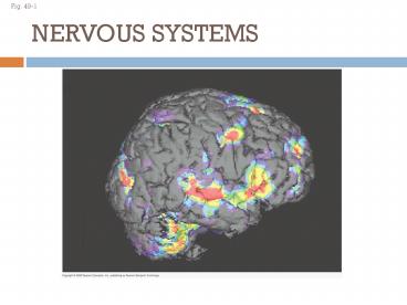
Fig. 49-1
2
Nervous systems consist of circuits of neurons
and supporting cells
- The simplest animals with nervous systems, the
cnidarians, have neurons arranged in nerve nets - A nerve net is a series of interconnected nerve
cells - More complex animals have nerves
3
- Nerves are bundles that consist of the axons of
multiple nerve cells - Sea stars have a nerve net in each arm connected
by radial nerves to a central nerve ring
4
Fig. 49-2a
Radial nerve
Nerve ring
Nerve net
(a) Hydra (cnidarian)
(b) Sea star (echinoderm)
5
- Bilaterally symmetrical animals exhibit
cephalization - Cephalization is the clustering of sensory organs
at the front end of the body - Flatworms show cephalization, with a small brain
and longitudinal nerve cord. They have the
simplest clearly defined Central Nervous System
(CNS).
6
- Annelids and arthropods have segmentally arranged
clusters of neurons called ganglia and a ventral
nerve cord.
7
Fig. 49-2b
Eyespot
Brain
Brain
Nerve cords
Ventral nerve cord
Transverse nerve
Segmental ganglia
(c) Planarian (flatworm)
(d) Leech (annelid)
8
Fig. 49-2c
Brain
Ganglia
Anterior nerve ring
Ventral nerve cord
Longitudinal nerve cords
Segmental ganglia
(e) Insect (arthropod)
(f) Chiton (mollusc)
9
- Nervous system organization usually correlates
with lifestyle - Sessile molluscs (e.g., clams and chitons) have
simple systems, whereas more complex molluscs
(e.g., octopuses and squids) have more
sophisticated systems
10
Fig. 49-2d
Brain
Spinal cord (dorsal nerve cord)
Brain
Sensory ganglia
Ganglia
(g) Squid (mollusc)
(h) Salamander (vertebrate)
11
- In vertebrates
- The Central Nervous System is composed of the
brain and spinal cord - The peripheral nervous system (PNS) is composed
of nerves and ganglia - Vertebrates have a hollow dorsal nerve cord.
12
Fig. 49-4
Peripheral nervous system (PNS)
Central nervous system (CNS)
Brain
Cranial nerves
Spinal cord
Ganglia outside CNS
Spinal nerves
13
Organization of the Vertebrate Nervous System
- The spinal cord conveys information from the
brain to the PNS - The spinal cord also produces reflexes
independently of the brain - A reflex is the bodys automatic response to a
stimulus - Examples Jerking your finger off a flame
- NOTE Conscious thought is not required in a
reflex.
14
- Stimulus detected by a receptor in the skin,
conveyed via a sensory neuron to an interneuron
in the spinal cord, which synapses with a motor
neuron, which will cause the effector, a muscle
cell to contract.
15
- The central canal of the spinal cord and the
ventricles of the brain are hollow and filled
with cerebrospinal fluid - The cerebrospinal fluid is filtered from blood
and functions to cushion the brain and spinal
cord - The cerebrospinal fluid also baths cells with
nutrients and carries away wastes.
16
Fig. 49-5
Gray matter
White matter
Ventricles
17
- The brain and spinal cord contain
- Gray matter, which consists of neuron cell
bodies, dendrites, and unmyelinated axons - White matter, which consists of bundles of
myelinated axons
18
Glia are cells that support neurons.
- Glia have numerous functions
- Astrocytes provide structural support for
neurons, regulate extracellular ions and
neurotransmitters, and induce the formation of a
blood-brain barrier that regulates the chemical
environment of the CNS - Oligodendrocytes form myelin sheaths in the
Central Nervous System - Schwann cells form myelin sheaths in the
Peripheral Nervous System.
19
Fig. 49-6
CNS
PNS
Neuron
VENTRICLE
Astrocyte
Ependy- mal cell
Oligodendrocyte
Schwann cells
Microglial cell
Capillary
(a) Glia in vertebrates
50 µm
(b) Astrocytes (LM)
20
The Peripheral Nervous System
- The PNS transmits information to and from the CNS
and regulates movement and the internal
environment - In the PNS, afferent neurons transmit information
to the CNS and efferent neurons transmit
information away from the CNS - Cranial nerves originate in the brain and mostly
terminate in organs of the head and upper body - Spinal nerves originate in the spinal cord and
extend to parts of the body below the head
21
- The Peripheral Nervous System is divided into
- The Motor (somatic) Nervous System, which carries
signals to skeletal muscles. It is a voluntary
system. - The Autonomic Nervous System, which regulates the
primarily autonomic visceral functions of smooth
and cardiac muscle. This is the involuntary
system.
22
Fig. 49-7-2
PNS
Efferent neurons
Afferent (sensory) neurons
Motor system
Autonomic nervous system
Hearing
Enteric division
Sympathetic division
Parasympathetic division
Locomotion
Hormone action
Circulation
Gas exchange
Digestion
23
- The autonomic nervous system transmits signals
that regulate the internal environment by
controlling smooth muscle and cardiac muscles,
including those in the gastrointestinal,
cardiovascular, excretory, and endocrine systems. - The autonomic nervous system has sympathetic,
parasympathetic, and enteric divisions
24
- The sympathetic division correlates with the
fight-or-flight response, when activated causes
the heart to beat faster and adrenaline to be
secreted. - The parasympathetic division promotes a return to
rest and digest - The enteric division controls activity of the
digestive tract, pancreas, and gallbladder
25
Fig. 49-8
Parasympathetic division
Sympathetic division
Action on target organs
Action on target organs
Dilates pupil of eye
Constricts pupil of eye
Inhibits salivary gland secretion
Stimulates salivary gland secretion
Sympathetic ganglia
Constricts bronchi in lungs
Relaxes bronchi in lungs
Cervical
Slows heart
Accelerates heart
Stimulates activity of stomach and intestines
Inhibits activity of stomach and intestines
Thoracic
Inhibits activity of pancreas
Stimulates activity of pancreas
Stimulates glucose release from liver inhibits
gallbladder
Stimulates gallbladder
Lumbar
Stimulates adrenal medulla
Promotes emptying of bladder
Inhibits emptying of bladder
Sacral
Promotes ejaculation and vaginal contractions
Promotes erection of genitals
Synapse
26
Fig. 49-9c
Cerebrum (includes cerebral cortex, white
matter, basal nuclei)
Diencephalon (thalamus, hypothalamus, epithalamus)
Midbrain (part of brainstem)
Pons (part of brainstem), cerebellum
Medulla oblongata (part of brainstem)
Diencephalon
Cerebrum
Hypothalamus
Thalamus
Pineal gland (part of epithalamus)
Brainstem
Midbrain
Pons
Pituitary gland
Medulla oblongata
Spinal cord
Cerebellum
Central canal
(c) Adult
27
Fig. 49-UN5
Cerebral cortex
Cerebrum
Thalamus
Forebrain
Hypothalamus
Pituitary gland
Midbrain
Pons
Spinal cord
Medulla oblongata
Hindbrain
Cerebellum
28
Fig. 49-UN1
29
The Brainstem
- The brainstem coordinates and conducts
information between brain centers - The brainstem has three parts the midbrain, the
pons, and the medulla oblongata
30
- The midbrain contains centers for receipt and
integration of sensory information - The pons regulates breathing centers in the
medulla - The medulla oblongata contains centers that
control several functions including breathing,
cardiovascular activity, swallowing, vomiting,
and digestion
31
Arousal and Sleep
- The brainstem and cerebrum control arousal and
sleep - The core of the brainstem has a diffuse network
of neurons called the reticular formation - This regulates the amount and type of information
that reaches the cerebral cortex and affects
alertness - The hormone melatonin is released by the pineal
gland and plays a role in bird and mammal sleep
cycles
32
Fig. 49-10
Eye
Input from nerves of ears
Reticular formation
Input from touch, pain, and temperature receptors
33
The Cerebellum
- The cerebellum is important for coordination and
error checking during motor, perceptual, and
cognitive functions - It is also involved in learning and remembering
motor skills
34
Fig. 49-UN2
35
The Diencephalon
- The diencephalon includes the thalamus, and
hypothalamus - The thalamus is the main input center for sensory
information to the cerebrum and the main output
center for motor information leaving the cerebrum - The hypothalamus regulates homeostasis and basic
survival behaviors such as feeding, fighting,
fleeing, and reproducing, thermostat, thirst, and
circadian rhythms
36
Fig. 49-UN3
37
The Cerebrum
- The cerebrum has right and left cerebral
hemispheres - Each cerebral hemisphere consists of a cerebral
cortex (gray matter) overlying white matter. - In humans, the cerebral cortex is the largest and
most complex part of the brain
38
Fig. 49-UN4
39
- A thick band of axons called the corpus callosum
provides communication between the right and left
cerebral cortices - The right half of the cerebral cortex controls
the left side of the body, and vice versa
40
Lateralization of Cortical Function
- The corpus callosum transmits information between
the two cerebral hemispheres - The left hemisphere is more adept at language,
math, logic, and processing of serial sequences - The right hemisphere is stronger at pattern
recognition, nonverbal thinking, and emotional
processing
41
Fig. 49-13
Right cerebral hemisphere
Left cerebral hemisphere
Thalamus
Corpus callosum
Basal nuclei
Cerebral cortex
42
The cerebral cortex controls voluntary movement
and cognitive functions
- Each side of the cerebral cortex has four lobes
frontal, temporal, occipital, and parietal - Each lobe contains primary sensory areas and
association areas where information is integrated
43
Fig. 49-15
Frontal lobe
Parietal lobe
Somatosensory cortex
Motor cortex
Somatosensory association area
Speech
Frontal association area
Taste
Reading
Speech
Hearing
Visual association area
Smell
Auditory association area
Vision
Temporal lobe
Occipital lobe
44
Emotions
- Emotions are generated and experienced by the
limbic system and other parts of the brain
including the sensory areas - The limbic system is a ring of structures around
the brainstem that includes the amygdala,
hippocampus, and parts of the thalamus - The amygdala is located in the temporal lobe and
helps store an emotional experience as an
emotional memory
45
Fig. 49-18
Thalamus
Hypothalamus
Prefrontal cortex
Olfactory bulb
Amygdala
Hippocampus
46
Consciousness
- Modern brain-imaging techniques suggest that
consciousness is an emergent property of the
brain based on activity in many areas of the
cortex
47
Memory and Learning
- Learning can occur when neurons make new
connections or when the strength of existing
neural connections changes - Short-term memory is accessed via the hippocampus
- The hippocampus also plays a role in forming
long-term memory, which is stored in the cerebral
cortex
Recommended
CrystalGraphics Presentations


