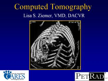
Computed Tomography - PowerPoint PPT Presentation
1 / 87
Title: Computed Tomography
1
Computed Tomography
- Lisa S. Ziemer, VMD, DACVR
2
Goals
- What is CT?
- CT capabilities
- Indications for referral
3
Computed Tomography
- Generates cross sectional images
- X-Rays are used for image collection
- Computer generates the images using data
collected during the study
4
Computed TomographyBasics
5
CARES CT Inside
6
Computed Tomography
7
Computed Tomography
8
Computed Tomography
9
CT vs. Radiography
- Eliminates superimposition, allowing detection of
lesions not visible on radiographs - CT discriminates 2000 densities
- Radiography discriminates only five
- 3-D reconstruction is possible
10
Viewing Images
Bone Window
Brain Window
CT gives you the ability to adjust the
brightness and contrast of the images, which
allows assessment of soft tissue and bone on the
same images.
11
Iodinated contrast material in CT
- Intravenous iodinated contrast medium is commonly
used in CT - Increased uptake in highly vascular tissue
neoplasia, inflammation - Prolonged uptake in neoplasia or other areas with
leaky vessels - Also used for angiography, intravenous urography
12
CT of the head
- Brain
- Nose
- Temporomandibular joints
- Middle ear
- Retrobulbar space
13
CT Brain
- Indications
- Seizures
- Behavior changes
- Other neurologic deficits
- Differentials
- Congenital defects
- Trauma
- Inflammation
- Neoplasia
- Infarction
14
Normal Brain
15
Hydrocephalus
- Obstructive
- Congenital
- Acquired
- Non-obstructive
- Ex Vacuo
16
Cerebrovascular Accident (CVA)
- Infarction
- Septic thromboembolism
- Neoplasia
- Coagulopathy
- Heartworm
- Idiopathic
- Infarction can be detected as early as 3- 6 hours
after onset. Enhancement within 24- 48 hours - Cerebral Hemorrhage
- Hypertension
- Neoplasia
- Vasculitis
- Coagulopathy
- CT extremely sensitive for acute hemorrhage.
Enhancement 6 days- 6 weeks.
17
Inflammatory Brain Disease
- Infectious
- Fungal
- Bacterial
- Protozoal (Neospora, Toxo)
- Viral (Distemper, FIV, FIP)
- Non-Infectious
- Breed Specific Encephalitides (Yorkshire,
Maltese, Pug) - Granulomatous Meningoencephalitis
Granulomatous Meningoencephalitis
18
Brain Tumors
- Mass effect
- Falx Shift
- Ventricular Asymmetry
- Edema
- Contrast Enhancement
- Less sensitive than MRI for caudal fossa tumors
and low grade neoplasms with insufficient
contrast enhancement
Meningioma
19
Pituitary Gland
Normal
Macroadenoma
20
(No Transcript)
21
Choroid Plexus Papilloma
22
139916/3y/MC/Labrador
Brain Metastasis
Choroid Plexus Papilloma
23
Masticatory Muscle Atrophy
Trigeminal nerve sheath tumor
24
Trigeminal Nerve Sheath Tumor
25
CT Ears
- Indications
- Vestibular disease
- Chronic ear disease
- Differentials
- Otitis
- Neoplasia
- Polyps
26
Normal Canine Middle and Inner Ear
27
Normal Bullae
Feline
Canine
28
Otitis MediaRadiography- 25 false negatives
29
Otitis Media
Feline
Canine
30
Aural tumors
- External ear canal neoplasia
- Ceruminal gland adenomas
- Ceruminal gland carcinomas
- Squamous cell carcinomas
- Primary middle ear tumors are less common
31
Aural tumors
- External ear canal neoplasia
- Ceruminal gland adenomas
- Ceruminal gland carcinomas
- Squamous cell carcinomas
- Primary middle ear tumors are less common
32
Nasal CT
- Indications
- Nasal Discharge/ Epistaxis
- Sneezing
- Pre-rhinoscopy or surgery
- Differentials
- Rhinitis
- Neoplasia
- Fungal infection
- Foreign body
33
Normal Canine Nasal Cavity
34
Normal Canine Nasal Cavity
35
Rhinitis
36
Canine Nasal Neoplasia
37
Canine Nasal Neoplasia
38
Nasal Aspergillosis
39
CT Orbit
- Indications
- Exophthalmus
- Epiphora
- Differentials
- Neoplasia
- Abscess
- Granuloma
- Mucocoele
- Tear Duct Obstruction
40
Retrobulbar Masses
- Typically present with exophthalmus
- Very difficult to diagnose radiographically
- Ultrasound is often helpful
- CT is ideal for imaging retrobulbar space
- DDx
- Neoplasia (particularly LSA and nasal tumors)
- Abscess
- Granuloma
- Mucocoele
41
Retrobulbar Mass secondary to Nasal Neoplasia
42
CT Temporomandibular Joint
- Indications
- Difficulty chewing
- Unable to open/ close mouth
- Pain with chewing
- Differential Diagnoses
- Fractures
- Ankylosis
- Neoplasia
43
Normal Temporomandibular Joints
44
Retroglenoid and Mandibular Condyle Fracture
45
Fracture of the Retroglenoid Process
46
Ankylosis of the TMJ
47
CT Skull
- Indications
- Tumor margins
- Swelling
- Trauma
- Differentials
- Fractures
- Neoplasia
- Osteomyelitis
48
Skull Fractures
- Can be very difficult to accurately diagnose with
radiographs - CT allows assessment for hematoma formation
- Acute hemorrhage is hyperdense (bright) on CT and
is surrounded by vasogenic edema
49
Left Sided Facial Swelling- Multilobular Tumor of
Bone
50
Left Sided Facial SwellingPrimary Bone Tumor-
Feline
51
CT Appendicular Skeleton
- Indications
- Elbow dysplasia
- Fissures / Fractures
- Bone tumor margins
- Questionable radiographic findings
52
Elbow Dysplasia
- Fragmented Medial Coronoid Process
- Incomplete Ossification of the Humeral Condyle
- Osteochondritis Dissecans (OCD)
- Elbow Incongruities
- Ununited Medial Humeral Epicondyle
- Ununited Anconeal Process
53
Fragmented Medial Coronoid Process
- Most common cause of elbow lameness in young dogs
- May or may not be a separate fragment
- Abnormal shape of MCP
- Fissures
- Radial incisure abnormalities
- Difficult to diagnose
- Non-displaced fragments
- Non-opaque fragments
- CT allows definitive diagnosis
54
Fragmented Medial Coronoid Process
Left
Right
?
55
Medial Coronoid Process88 vs. 23 sensitivity
(CT vs. radiographs)
56
Image Reconstruction
Normal
FMCP
57
Osteochondritis dissecans of the medial humeral
condyle
- Clinical signs similar to FCP
- Etiology
- Heritable genetic disease.
- Failure of enchondral ossification, cartilage
fragmentation, subchondral sclerosis. - Likely influenced by high energy diets and rapid
growth. - Difficult to diagnose small lesion,
superimposition of olecranon
58
Humeral Condyle OCD
59
Incomplete Ossification of the Humeral Condyle
- Heritable condition in Spaniel breeds, rare in
others - Other forms of elbow dysplasia are rare in
Spaniels! - Two centers of ossification appear 22 days of
age, fuse by 84 days - Defect of bony fusion of medial and lateral
ossification centers - Difficult to diagnose radiographically
- Can lead to condylar fractures- 50 Y or T shaped
60
Incomplete Ossification of the Humeral Condyle
61
Opposite Limb
62
Questionable or subtle radiographic findings
63
CT Spine
- Indications
- Neurologic deficits
- Pain
- Differentials
- Fractures
- Disc disease / LS disease
- Neoplasia (vertebral, spinal, nerve root)
64
Thoracolumbar Disc ProlapsePost Myelography
Normal
IVD Prolapse
65
Intervertebral Disc Prolapse (Mineralized Disc)
66
Spinal Fractures
- Radiography is inadequate to rule out vertebral
fractures and subluxation in the acute canine
spinal trauma patient. Radiography cannot
reliably be used to detect compressive lesions.
CT, where available, is therefore recommended in
patients with a high clinical suspicion of such
injury. - Radiographs
- Fractures NPV 48
- Spinal cord instability NPV 35
- Sensitivity for all fractures 72
- Kinns, et al. Vet Rad and US, 2006.
67
Spinal Fractures
68
Lumbosacral Disease
- Lumbosacral Stenosis
- Congenital Malformation
- Inflammation
- Neoplasia
- Degenerative Changes
- Disc Herniation
- Ligamentous hypertrophy
- Vertebral DJD
- Spondylosis
- Instability
69
Lumbosacral Disease
NORMAL
L7- S1 DISC PROLAPSE
70
2- D Reconstruction
71
Vertebral Tumor (Metastatic OSA)
72
Peripheral nerve sheath tumors
- Nerve sheath tumors typically present with pain,
neurologic deficits, muscle atrophy - Very difficult to diagnose
- On CT may be very subtle thickening, or may be a
large mass with extension into the spinal canal
C7 nerve root tumor post myelogram
73
Brachial Plexus Tumor
74
Brachial Plexus Tumor
75
CT chest and abdomen
- Indications
- Mediastinum
- Neoplasia
- Cyst
- Abscess/ granuloma
- Lung interstitium
- Metastatic disease
- Primary tumors
- Lung lobe torsion
- Pleural effusion
- Thoracic Radiographs are indeterminate
- May change diagnosis in more than 50 of cases
- Liver / kidney Vascular anatomy
- Mass location and margins
76
CT chest and abdomen
- Indications
- Mediastinum
- Neoplasia
- Cyst
- Abscess/ granuloma
- Lung interstitium
- Metastatic disease
- Primary tumors
- Lung lobe torsion
- Pleural effusion
- Thoracic Radiographs are indeterminate
- May change diagnosis in more than 50 of cases
- Liver / kidney Vascular anatomy
- Mass location and margins
77
Normal Lung
78
Pulmonary Metastatic Disease
- CT allows detection of nodules as small as 1 mm
- Pulmonary nodules not visible on thoracic
radiographs until at least 7 mm - Only 9 of metastases visible on CT were visible
on radiographs (Nemanic, et al, JVIM, 2006)
79
Primary pulmonary tumor with metastasis to
tracheobronchial LNs
- Primary pulmonary tumors commonly metastasize to
the tracheobronchial lymph nodes - CT 83 sensitivity and 100 specificity
(Paolini, et al. JAVMA, 2006) - Radiography did not detect TBLN enlargement in
this paper
80
Idiopathic Pulmonary Fibrosis of the West
Highland White Terrier
81
Malignant Effusion/ Cavitated Pulmonary Mass
82
Pulmonary Bulla
83
Tumor margins
- CT is extremely useful for surgical planning
- Identify margins
- Determine whether a tumor is resectable/ assess
for invasion into adjacent structures
84
Conclusions
- CT is a very sensitive tool for a wide variety of
organs and lesions. - Best established head, elbows, spine.
- Increasing new clinical applications.
- Compared To MRI
- CT is excellent for bone lesions, nasal lesions
- Good for structural or mass lesions in the brain
- MRI is better for non-structural brain lesions
- With CT, only axial plane acquired (3 planes can
be acquired with MRI)
85
How do you get this done?
- For outpatient studies, call CARES and make an
appointment for CT. Patient dropped off through
the emergency service return home on the same
day. - For ongoing care or further assessment, make an
appointment with a specialist (oncology, surgery,
internal medicine, ophthalmology). - Recent CBC/ CS (within 14 days) may be obtained
by you or at CARES. - CT reports will be faxed to your office the same
day. - Feel free to call any time with questions!
86
(No Transcript)
87
Acknowledgements
- PetRad
- Rob McLear
- CARES
- BJ DiTullio
- Donna Steckley
- Jon Rappaport
- Bob Cohen


![[PDF] READ] Free Cardiovascular Computed Tomography (Oxford Specialist PowerPoint PPT Presentation](https://s3.amazonaws.com/images.powershow.com/10075957.th0.jpg?_=202407100710)
![[PDF] READ] Free Cone Beam Computed Tomography: Oral and Maxillofacial PowerPoint PPT Presentation](https://s3.amazonaws.com/images.powershow.com/10076634.th0.jpg?_=202407110310)

![get [PDF] DOWNLOAD Cone Beam Computed Tomography in Endodont PowerPoint PPT Presentation](https://s3.amazonaws.com/images.powershow.com/10084529.th0.jpg?_=20240724058)
