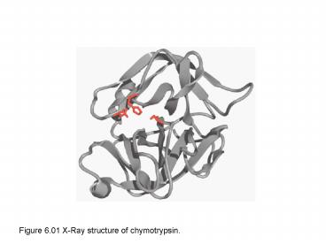
Figure 6'01 XRay structure of chymotrypsin' PowerPoint PPT Presentation
Title: Figure 6'01 XRay structure of chymotrypsin'
1
Figure 6.01 X-Ray structure of chymotrypsin.
2
Figure 6.02 Reaction coordinate diagram for the
reaction AB C
A BC
3
Figure 6.03 Reaction coordinate diagram for a
reaction in which reactants and products have
different free energies.
4
Figure 6.04 Effect of a catalyst on a chemical
reaction.
5
Figure 6.05 Amino acid side chains that can act
as acid-base catalysts.
6
Figure 6.06 Reaction coordinate diagram for a
reaction accelerated by covalent catalysis.
7
Figure 6.07 Protein groups that can act as
covalent catalysts.
8
Figure 6.08 The catalytic triad of chymotrypsin.
9
Figure 6.09 The catalytic mechanism of
chymotrypsin and other serine proteases.
10
Figure 6.11 Effect of very tight substrate
binding on enzyme catalysis.
11
Figure 6.12 Transition state stabilization in the
osyanion hole.
12
Figure 6.13 Formation of a low-barrier hydrogen
bond during catalysis in chymotrypsin.
13
Figure 6.14 Proximity and orientation effect in
catalysis.
14
Figure 6.18 Specificity pockets of three serine
proteases.
15
Figure 6.19 Activation of chymotrypsinogen.
PowerShow.com is a leading presentation sharing website. It has millions of presentations already uploaded and available with 1,000s more being uploaded by its users every day. Whatever your area of interest, here you’ll be able to find and view presentations you’ll love and possibly download. And, best of all, it is completely free and easy to use.
You might even have a presentation you’d like to share with others. If so, just upload it to PowerShow.com. We’ll convert it to an HTML5 slideshow that includes all the media types you’ve already added: audio, video, music, pictures, animations and transition effects. Then you can share it with your target audience as well as PowerShow.com’s millions of monthly visitors. And, again, it’s all free.
About the Developers
PowerShow.com is brought to you by CrystalGraphics, the award-winning developer and market-leading publisher of rich-media enhancement products for presentations. Our product offerings include millions of PowerPoint templates, diagrams, animated 3D characters and more.

