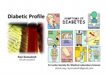
Diabetic Profile - PowerPoint PPT Presentation
Title:
Diabetic Profile
Description:
Is group of tests that are used to diagnose diabetes or its complications , it includes: Blood glucose 4 types: FBS, PPBS, RBS, OGGT Urine Analysis Urine Sugar / Urine Protein /Urine Microalbumin / Ketones HbA1C Insulin ICA (islent cell antibody) for type I C-peptide – PowerPoint PPT presentation
Number of Views:1719
Learn more at:
http://slsmls.org
Title: Diabetic Profile
1
Diabetic Profile
Ravi Kumudesh MSc/BSc/Dip(MLT)
Sri Lanka Society for Medical Laboratory
Science slsmls.org / kumudeshr_at_gmail.com
2
Diabetes Mellitus
- It is a chronic disease due to disorder of
carbohydrate metabolism, due to insulin
deficiency results in hyperglycemia (increased
blood glucose level) glucourea (presence of
glucose in urine). - Associated with several changes in metabolism
such as metabolism of proteins fats.
3
Clinical Biochemical Findings in Diabetes
- Glucosuria.
- Large volume of urine
- increase urination frequency (Polyuria)
- Polyphagia (eats more frequently)
- Several metabolic changes
4
Metabolic changes in diabetes
- Include increase in
- Fat catabolisim leads to increase in FFAs in
blood liver. - Acetyl.coA leads to increase formation of
cholesterol risk of atherosclerosis. - ketone bodies generation in blood and urine leads
to acidosis. - catabolism of tissue protein due to energy
requirement (because glucose can't uptake by
cells) lead to weight loss and increase in level
of amino acids in blood more formation of urea
by deamination of amino acid.
5
Types of diabetes
- Type I diabetes mellitus (TIDM)
- Type 2 diabetes mellitus (TIIDM)
- Gestational diabetes mellitus (GDM)
- Other "due to drugs or chemicals"
6
Diabetic profile
- Is group of tests that are used to diagnose
diabetes or its complications , it includes - Blood glucose
- 4 types FBS, PPBS, RBS, OGGT
- Urine Analysis
- Urine Sugar / Urine Protein /Urine Microalbumin /
Ketones - HbA1C
- Insulin
- ICA (islent cell antibody) for type I
- C-peptide
7
Urine Analysis
8
1. Urine Sugar
Detection of urinary glucose (Glucosuria)
9
Glucosuria
- First-line screening test for diabetes mellitus
- Normally glucose does not appear in urine until
the plasma glucose rises above 160-180 mg/dl. - In certain individuals due to low renal threshold
glucose may be present despite normal blood
glucose levels. - Conversely renal threshold increases with age so
many diabetics may not have Glycosuria despite
high blood sugar levels.
Positive Benedicts test
10
- A specific and convenient method to detect
glucosuria is the paper strip impregnated with
glucose oxidase and a chromogen system
(Clinistix, Diastix), which is sensitive to as
little as 0.1 glucose in urine. - Diastix can be directly applied to the urinary
stream, and differing color responses of the
indicator strip reflect glucose concentration. - Benedicts and Fehlings test can also detect
glucosuria.
Diastix- Reagent strips
11
Microalbuminuria
2. Urine Microalbumin
12
Microalbuminuria
- The importance of micro- albuminuria in the
diabetic patient is that it is a signal of early
reversible renal damage. - Performing an albumin-to-creatinine ratio is
probably easiest. - Microalbuminuria is a common finding (even at
diagnosis) in type 2 diabetes mellitus and is a
risk factor for macro vascular (especially
coronary heart) disease.
Gradation of turbidity is linked to protein
concentration
13
Microalbuminuria
- May be defined as an albumin excretion rate
intermediate between normality (2.5-25 mg/day)
and macroalbuminuria (250mg/day). - The small increase in urinary albumin excretion
is not detected by simple albumin stick tests and
requires confirmation by careful quantization in
a 24 hr urine specimen.
14
Assays for Microalbuminurea
Qualitative
- Dipstick method
Quantitative
- Enzyme linked Immunosorbant assay
- Radioimmuno assay
- Immunoturbidometric assay
15
Specimen Collection for Microalbimin
- Collect freshly voided urine in a clean, dry
container - Preservatives should be avoided
- Samples which cannot be tested within 3 days of
collection should be refrigerated - Samples should not be frozen
- The test should be free from significant
interference from glucosuria, pH, ketonuria or
bacterial contamination
16
MICRAL Strips
- Micral strip screening tests offer a
cost-effective method of screening - Dip sticks show acceptable sensitivity (95) and
specificity (93) - All positive tests should be confirmed by more
specific methods
17
False Positives
- Hyper filtration (Newly diagnosed diabetes)
- Exercise
- Marked hypertension
- Congestive Heart Failure
- Urinary Tract Infection
- Acute febrile illness
18
3. Urine Ketone Bodies
Ketonuria
19
What Are Ketones?
- Acids that result when the body does not have
enough insulin and uses fats for energy - May occur when insulin is not given, during
illness or extreme bodily stress, or with
dehydration - Can cause abdominal pain, nausea, and vomiting
- Without sufficient insulin ketones continue to
build up in the blood and result in diabetic
ketoacidosis (DKA)
20
Why Test for Ketones?
- DKA is a critical emergency state
- Early detection and treatment of ketones prevents
diabetic ketoacidosis (DKA) and hospitalizations
due to DKA - Untreated, progression to DKA may lead to severe
dehydration, coma, permanent brain damage, or
death - DKA is the number one reason for hospitalizing
children with diabetes
21
When Should Ketones Be Checked?
- The DMMP should specify, generally
- When blood glucose remains elevated
- During acute illness, infection or fever
- Whenever symptoms of DKA are present
- Nausea
- Vomiting or diarrhea
- Abdominal Pain
- Fruity breath odor
- Rapid breathing
- Thirst and frequent urination
- Fatigue or lethargy
- Common symptoms including fruity odor to breath,
nausea, vomiting, drowsiness, abdominal pain
22
How Quickly Does DKA Progress?
- An isolated high blood glucose reading, in the
absence of other symptoms is not cause for alarm - DKA usually develops over hours, or even days
- DKA can progress much more quickly for students
who use insulin pumps, or those who have an
illness or infection - Most at risk when symptoms of DKA are mistaken
for flu and high blood glucose is unchecked and
untreated
23
Checking for Ketones
- Urine testing
- Most widely used method
- Blood testing
- Requires a special meter and strip
- Procedure similar to blood glucose checks
24
How to Test Urine Ketones
- 1. Gather supplies
- 2. Student urinates in clean cup
- 3. Put on gloves, if performed by someone other
than student - 4. Dip the ketone test strip in the cup
containing urine. Shake off excess urine - 5. Wait 15 - 60 seconds
- 6. Read results at designated time
- 7. Record results, take action per DMMP
25
Test Results Color Code
- no ketones
- trace
- small
- moderate
- large ketones present
26
Considerations
- Colors on strips and timing vary according to
brand - If using a scale with urine glucose and urine
ketones, be sure to read the correct scale when
testing for ketones - Follow package instructions regarding expiration
dates, time since opening, correct handling,
etc., as incorrect results may occur
27
How To Test for Blood Ketones
- Prepare lancing device
- Wash hands using warm soapy water and dry them
completely - Remove the test strip from its foil packet
- Insert the three black lines at the end of the
test strip into the strip port - Push the test strip in until it stops
28
How To Test for Blood Ketones
- Touch the blood drop to the purple area on the
top of the test strip. The blood is drawn into
the test strip - Continue to touch the blood drop to the purple
area on the top of the test strip until the
monitor begins the test - The blood ß-Ketone result shows on the display
window with the word KETONE
29
Ketonuria
- Qualitative detection of ketone bodies can be
accomplished by nitroprusside tests (Acetest or
Ketostix), Rotheras test etc. - These tests do not detect Beta-hydroxy butyric
acid, which lacks a ketone group - Ketone bodies may be present in a normal subject
as a result of simple prolonged fasting.
Ketostix- Reagent strips
Positive Rotheras test
30
Blood Glucose Levels
31
1. Fasting blood sugar (FBS)
- Measures blood glucose after fasting for at least
8-12 hrs - It often is the first test done to check for
diabetes. - patient with mild or borderline diabetes may
present with normal FBG values. - If diabetes is suspected, GTT can confirm the
diagnosis. - Normal levels
- 70-110mg/dl
32
2. Post-Prandial Blood Sugar (2-hour PPBS)
- After the patient fasts for 12 hours, a meal is
given which contains starch and sugar (approx.
100 gm). - Then after 2 hours blood is collected to measure
glucose level. - home blood sugar test is the most common way to
check 2-hour postprandial blood sugar levels.
33
3. Random blood sugar (RBS)
- measures blood glucose randomly at any time
throughout the day without patient fasting. - it is useful because glucose levels in healthy
people dont vary widely throughout the day. - blood glucose levels that vary widely may
indicate a problem.
34
4. Oral glucose tolerance test (OGTT)
- Glucose Tolerance is defined as the capacity of
the body to tolerate an extra load of glucose or
it measures the body's ability to use glucose. - It is series of blood glucose measurements taken
after drink glucose liquid - It is considered as definitive diagnostic test
for DM. - It is ordered to
- Confirm the diagnosis, in pre-diabetic
- Diagnose gestational diabetes (most commonly)
- Recommended if 100-126 mg/dL (5.5 mmol/L-7.0
mmol/L)
35
Procedure
- Arrive FBS After an overnight fasting
- (10-12 hrs)
- Drink 75-100g dissolved in 250-300ml of water
and given orally. - After drink blood samples and urine are
collected every 30min for 3hrs - (1 hr, 1.5 hr , 2hr, 2.5hr, 3hr )
- A curve between time and blood glucose
concentration, is plotted.
36
Other types of OGTT
- Extended GTT
- Glucose measured for 4-5 hrs after giving
glucose to see how the curve behaves below the
normal fasting glucose limits. Done in some
conditions causing hypoglycaemia. - Cortisone Stressed GTT Can be used for
detecting latent DM. - Intravenous GTT
- Is done if oral glucose is not tolerated or oral
GTT curve is flat. - In these cases 20 glucose as 0.5g glucose/Kg
body weight. - Usually peak occurs within 30 min after infusion
and returns to normal after 90 min.
37
Interpretation
- Normal Response
- FBS is normal. After 1 hr it will rise, returns
to normal fasting level within 2 hours. - Diabetic curve
- FBS 140mg/dl or 7.8 mmol/L. After 2 hr
200mg/dl (11 mmol/L) or more. Glucosuria is
usually seen - Impaired GTT
- with 2hrs glucose level between 140mg/dl -
200mg/dl - It is not abnormal but must be followed up for
DM.
38
Interpretation
- Renal Glycosuria
- Curve is normal due to lowered renal threshold
one or more samples of urine contain glucose. - Lag storage/Alimentary Type
- FBS is normal. Due to rapid absorption, maximum
level is found at 30 min (180mg/dl). glucosuria
is seen hypoglycaemic levels may be reached at
end of 2 hours. - Flat curve of enhanced glucose tolerance
- FBS is normal. Throughout the test the level
does not vary 20mg.
39
Hypoglycemia
- When blood glucose falls below 60 mg/dl.
- Causes
- Most commonly seen in overdose of insulin in
treatment of DM. - Hypothroidism.
- Insulin secreting tumours of pancrease rare.
- Hypoadrenahsm (Addison's disease)
- Hypopitruitism.
- Severe exercise.
- Starvation.
40
Measuring glucose level
- Principle
Glucose H2O O2 Gluconic
acid H2O2 2H2O2 4 aminoantipyrine PHBS
Quinoneimine dye H2O
Red color
GOD
POD
41
Kit components
- Glucose Oxidase Reagent
- mixture of
- glucose oxidase peroxidase
aminoantipyrine buffer - Glucose standard Reagent
- conc. 100mg/dl or 5.55 mmol/L
42
Procedure
- Prepare the reaction as the following
- Mix, incubates at 37oC for 10min
- Read abs at 510nm
Sample Standard Reagent blank
1ml 1 ml 1 ml Glucose oxidase reagent
10 µl - Sample (serum)
- 10 µl - Glucose standard
43
Calculation
Glucose conc. Abs. Sample X Conc.
Standard Abs. Standard ..
44
Bedside Method
45
Special Tests
46
1. GlycohaemoglobinHb A1C
47
Hb A1C
- HbA1C is glucose bound to hemoglobin
- Measures blood glucose conc. over a longer period
of time - It indicates how well diabetes has been
controlled in the 2-3 months before the test. - The A1C level is directly related to
complications from diabetes - Type of sample whole blood in EDTA tube
- Normal Values
- Glycohemoglobin A1c4.5-5.7
- Total glycohemoglobin5.3-7.5
48
Key Messages
- Glycated hemoglobin (A1C) ? measure every 3
months (6 months if stable at target) - Self monitoring Blood Glucose (SMBG) is an aid to
assess interventions and hypoglycemia - Individualize the frequency of SMBG
- SMBG and continuous glucose monitoring (CGM)
needs to be linked with structured educational
program to facilitate behavior change
49
Glycated Hemoglobin A1C
- Reliable measure of mean plasma glucose over 3-4
months - Valuable indicator of treatment effectiveness
- Measure every 3 months when glycemic targets are
not being met or treatments adjusted - Measure every 6 months if stable at glycemic
targets
50
Conditions that can Affect Value
Factors affecting A1C Increased A1C Decreased A1C Variable Change in A1C
Erythropoiesis B12/Fe deficiency Decreased erythropoiesis Use of EPO, Fe, or B12 Reticulocytosis Chronic liver Dx
Altered hemoglobin Fetal hemoglobin Hemoglobinopathies Methemoglobin
Altered glycation Chronic renal failure ??erythrocyte pH ASA, vitamin C/E Hemoglobinopathies ? erythrocyte pH
Erythrocyte destruction Splenectomy Hemoglobinopathies Chronic renal failure Splenomegaly Rheumatoid arthritis HAART meds,
Assays Hyperbilirubinemia Carbamylated Hb ETOH Chronic opiates Hypertriglyceridemia
51
2. Insulin Levels
52
Insulin Test - Clinical Relevance
- Insulin is the primary hormone responsible for
controlling glucose metabolism, and its secretion
is governed by plasma glucose concentration. - The insulin molecule is synthesized in the
pancreas - The principal function of insulin is to control
the uptake and utilization of glucose in the
peripheral tissues. - Insulin concentrations are severely reduced in
insulindependent diabetes mellitus (IDDM) Other
conditions, non-insulin-dependent diabetes
mellitus (NIDDM), obesity, and some endocrine
dysfunctions.
53
Insulin - Test Principle
- The Insulin ELISA is a two-site enzyme
immunoassay utilizing the direct sandwich
technique with two monoclonal antibodies directed
against separate antigenic determinants of the
insulin molecule. - Specimen, control, or standard is pipetted into
the sample well, then followed by the addition of
peroxidase-conjugated anti-insulin antibodies. - Insulin present in the sample will bind to
anti-insulin antibodies bound to the sample well,
while the peroxidase-conjugated anti-insulin
antibodies will also bind to the insulin at the
same time. - After washing to remove unbound enzyme-labelled
antibodies, TMB-labelled substrate is added and
binds to the conjugated antibodies. - Acid is added to the sample well to stop the
reaction, and the colorimetric endpoint is read
on a microplate spectrophotometer set to the
appropriate light wavelength.
54
3. Islet Cell Antibody (ICA)
55
Demonstration of Islet Cell Antibody (ICA)
- Using the indirect fluorescent antibody method
enables serologic assessment or possible
detection of pancreatic disease. - The presence of a (histologically defined)
circulating antibody to one or more of the islet
cell antigens can aid in patient diagnosis and
prognosis. - The substrate utilized in this kit is sections of
monkey pancreas. Islet Cell antibodies have been
associated with a group of "autoimmune" endocrine
disorders, more specifically with insulin
dependent diabetes. - Organ-specific autoimmunity is characterized by
the presence of antibodies in patients that can
be detected years before the onset of the
clinical symptoms. - Patients with autoimmune thyroiditis, adrenalitis
or gastritis have an increased risk of developing
insulin dependent diabetes at any age.
56
Test Principle and Procedure
- The indirect fluorescent antibody test is used
for the detection of human IgG antibody to the
antigens of monkey pancreas islet cells. - Tissue is placed in the wells of specially
prepared microscope slides. - Dilutions of patient sera are placed on the wells
where antibody, if present, binds to the antigen.
- The reaction is visualized through the use of a
conjugate. - The conjugate is fluorescein isothiocyanate
(FITC) labeled, anti-human IgG - Excitation of the FITC by ultraviolet (UV) light
causes this dye to emit longer, visible,
wavelengths of light in the yellow-green portion
of the color spectrum. - The conjugate will bind with human IgG antibodies
attached to the antigens causing fluorescence
when viewed through a microscope equipped with a
UV light source
57
4. C-peptide Test
58
C-peptide Test
- C-peptide testing can be used for a few different
purposes - C-peptide is a substance produced by the beta
cells in the pancreas when pro insulin splits
apart and forms one molecule of C-peptide and one
molecule of insulin - Insulin is the hormone that is vital for the body
to use its main energy source, glucose - Since C-peptide and insulin are produced at the
same rate, C-peptide is a useful marker of
insulin production
59
When is it ordered?
- Sweating
- Palpitations
- Hunger
- Confusion
- Blurred vision
- Fainting
- In severe cases, seizures
- loss of consciousness
60
What does the test result mean?
- A high level of C-peptide generally indicates a
high level of endogenous insulin production. This
may be in response to a high blood glucose caused
by glucose intake and/or insulin resistance. - A high level of C-peptide is also seen
with insulinomas and may be seen with low blood
potassium, Cushing syndrome, and renal failure - When used for monitoring, decreasing levels of
C-peptide in someone with an insulinoma indicate
a response to treatment levels that are
increasing may indicate a tumor recurrence - A low level of C-peptide is associated with a low
level of insulin production. This can occur when
insufficient insulin is being produced by
the beta cells, with diabetes for example, or
when production is suppressed by treatment
with exogenousinsulin
61
Not Only Test, But .?
62
Thank You !


