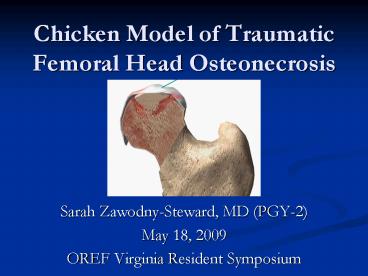
Chicken Model of Traumatic Femoral Head Osteonecrosis PowerPoint PPT Presentation
1 / 25
Title: Chicken Model of Traumatic Femoral Head Osteonecrosis
1
Chicken Model of Traumatic Femoral Head
Osteonecrosis
- Sarah Zawodny-Steward, MD (PGY-2)
- May 18, 2009
- OREF Virginia Resident Symposium
2
Osteonecrosis
- Ischemic death of cellular elements in the
epiphyses of bone - Deterioration of subchondral bone and loss of
mechanical integrity
3
Etiology
- No discrete agent or pathologic mechanism
identified, just theories - Altered blood flow
- Direct cellular toxicity
- Impaired mesenchymal cellular differentiation
- Factors
- Corticosteroids
- Coagulation abnormalities
- Alcohol use
- Genetic polymorphisms
- Trauma (dislocation or fracture)
4
Femoral Head Arterial Supply
- Proximal epiphyseal region is encased in
avascular cartilage - Increased risk of ischemia when periosteal and
endosteal blood supply is interrupted
5
Clinical Relevance
- Unrelenting course of pain and dysfunction
leading to femoral head collapse - Relatively young, active patients
- Mean age of presentation 38 years
- 20,000 new cases diagnosed annually
- Approximately 10 of all THAs
- Not optimal for young patients
- Conservative treatment options currently delay,
but do not halt progression - core decompression, osteotomy, nonvascularized
bone matrix grafting, free vascularized fibular
grafts, resurfacing procedures
6
Animal Model of Osteonecrosis
- Experimental techniques must mimic clinical
situations - Regardless of where circulation is initially
impeded (arteries, veins, capillaries,
sinusoids), the endpoint is interruption of
arterial flow - The lack of effective treatments for ON is mainly
due to the lack of an ideal animal model.
7
Why Chickens?
- Existing models
- Rat
- Piglet
- Emu
- Sheep
- Quadruped animal models have failed to produce
the human form of ON. - Majority do not progress to collapse of the
femoral head - Biomechanical environment of the hip joint in
bipedal animals is similar to humans. - Chickens treated with glucocorticoid steroids
have demonstrated the human form of ON in our
research laboratory (Dr. Cui et al.)
8
Traumatic Model of ON
- Surgical hip dislocation and disruption of the
vascular supply to the femoral head - Left hip is experimental side, Right serves as
control.
9
- Procedure
- 2 cm longitudinal incision over the greater
trochanter - Fascia of gluteus maximus incised to expose the
greater trochanter - Joint capsule detached and Ligamentum teres cut
- Femoral head dislocated from the acetabulum
10
Procedure
- Periosteum of the femoral neck scored and
surrounding periosteal vessels severed - Femoral head relocated
- Surgical incision closed in layers
Microfil (orange) injected into arterial system
of chickens to map vessels supplying the femoral
head.
11
Methods
- Total of 24 chickens
- 18wk female white leghorn
- 6 euthanized for tissue harvest at 4 time points
4, 8, 12, and 20 weeks - Femoral head and neck samples fixed in Formalin
- MicroCT
- Histological analysis
- Decalcification with 0.25M EDTA for 4-5 weeks
- Embedded in paraffin
- 7 µm coronal sections stained with HE
12
Quantitative µCT imaging analysis
13
(No Transcript)
14
(No Transcript)
15
Histology of Human Osteonecrosis
- Loss of thickness of trabecular bone
- Empty lacuna due to osteocyte death
- Appositional new bone formation on necrotic
trabeculae - Subchondral collapse, fracture, and/or cysts
- Marrow edema and/or necrosis
- Depletion of hematopoietic cells
- Fat cells are necrotic in the marrow
- Marrow space filled with eosinophilic fibrinoid
material
16
Control (10x)
8R (2x)
8L (2x)
Empty Lacunae
Normal Osteocytes
20 wk Experimental (10x)
17
20 wk (8L) at 40x Necrotic Bone
New appositional bone formation
Dead Osteocytes
18
Macroscopic Findings
Collapse
Subchondral Fracture
Necrotic Bone
Subchondral Cyst
Human specimen collected at time of Total Hip
Arthroplasty
19
20 week (10L) Subchondral cyst formation
20
20 week (9L) Collapse of subchondral bone, void
filled by fibrous tissue
21
Conclusions
- Histologic changes consistent with Osteonecrosis
seen as early as 4 weeks - Trabecular bone loss, empty osteocyte lacunae,
new bone formation on existing trabeculae - Progression to human form of disease
- Cyst formation and collapse by 20 weeks
- MicroCT analysis shows decreased bone volume and
increased porosity compared to controls. - Successful traumatic model of osteonecrosis can
be used to develop innovative treatments - Tissue Engineering
22
Core Decompression
- Most commonly used head preserving procedure for
pre-collapse osteonecrosis - Performed with or without bone graft or bone
graft substitute
Core decompression performed on chicken femoral
head a 1.6 mm k-wire and a 2 mm drill bit were
used for testing under the fluoroscopic guidance,
the femurs were then dissected to examine the
position of the core tracts.
23
Bone Graft Substitute
- Osteoconductive PLAGA sintered microsphere
scaffold - Osteoinductive and Angiogenic Stem cells
expressing BMP and VEGF concurrently
SEM images of D1 cells on the PLAGA sintered
microsphere scaffolds at day 21(C 80)
24
Special Thanks
- UVa Academic Orthopaedic Training Program
- Musculoskeletal Research Training Grant T32
AR0509601 from the NIAMS, NIH - Orthopaedic Research Education Foundation
- OREF Resident Clinician-Scientist Training Grant
- Orthopaedic Trauma Association
- OTA Resident Research Grant
25
References
- Mont MA, Jones LC, Hungerford DS. Nontraumatic
osteonecrosis of the femoral head ten years
later. J Bone Joint Surg Am 200688(5)1117-32.4.
- Cui Q, Wang GJ, Su CC, Balian G. The Otto Aufranc
Award. Lovastatin prevents steroid induced
adipogenesis and osteonecrosis. Clin Orthop Relat
Res 1997(344)8-19. - Troy KL, Lundberg HJ, Conzemius MG, Brown TD.
Habitual hip joint activity level of the penned
EMU (Dromaius novaehollandie). Iowa Orthop J
20072717-23. - Reed KL, Conzemius MG, Robinson RA, Brown TD.
Osteocyte-based image analysis for quantitation
of histologically apparent femoral head
osteonecrosis application to an emu model.
Comput Methods Biomech Biomed Engin
20047(1)25-32. - Reed KL, Brown TD, Conzemius MG. Focal cryogen
insults for inducing segmental osteonecrosis
computational and experimental assessments of
thermal fields. J Biomech 200336(9)1317-26. - Conzemius MG, Brown TD, Zhang Y, Robinson RA. A
new animal model of femoral head osteonecrosis
one that progresses to human-like mechanical
failure. J Orthop Res 200220(2)303-9. - Reed KL, Brown TD. Elastic modulus and strength
of emu cortical bone. Iowa Orthop J 20012153-7. - http//www.orthosupersite.com/view.asp?rID28890
- Koo KH, et al. Bone Marrow Edema and Associated
Pain in Early Stage Osteonecrosis of the Femoral
Head Prospective Study with Serial MR Images.
Radiology, Dec 1999 213 715 - 722. - McGrory BJ et al. Current Practices of AAHKS
Members in the Treatment of Adult Osteonecrosis
of the Femoral Head. J Bone Joint Surg Am.
2007891194-1204.

