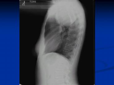
Evaluation of Mediastinal Mass PowerPoint PPT Presentation
1 / 24
Title: Evaluation of Mediastinal Mass
1
(No Transcript)
2
(No Transcript)
3
(No Transcript)
4
(No Transcript)
5
Evaluation of Mediastinal Mass
- Leslie Proctor, M.D.
- November 21, 2008
6
Mediastinal Anatomy
- Includes structures bound by
- the thoracic inlet
- diaphragm
- sternum
- vertebral bodies
- and pleura
- Has 3 compartments
- Anterior
- Middle
- Posterior
7
The differential diagnosis of a mediastinal mass
depends upon the anatomic compartment in which it
arises. Redrawn from Baue, AE, et al. Glenn's
Thoracic and Cardiovascular Surgery. 5th ed.
Appleton Lange, Norwalk, CT, 1991.
8
Mediastinal Anatomy
- Middle Compartment is bounded by
- Anterior Compartment includes
- The pericardium anteriorly
- The posterior pericardial reflection
- The diaphragm
- The thoracic inlet.
- This compartment includes the heart,
intrapericardial great vessels, pericardium, and
trachea.
- Thymus
- Extrapericardial aorta and its branches
- The great veins
- Lymphatic tissue.
Posterior Compartment
- Extends from the posterior pericardial
reflection to the posterior border of the
vertebral bodies and from the first rib to the
diaphragm. - It includes the esophagus, vagus nerves,
thoracic duct, sympathetic chain, and azygous
venous system
9
Anatomic Distribution of Masses
- Anterior Mediastinum
- Thymoma
- Thymic tumors and cysts
- Germ cell tumors
- Lymphomas
- Intrathoracic goiter and thyroid tumors
- Parathyroid adenomas
- Connective tissue tumors
- lipomas and liposarcomas
- lymphangiomas
- hemangiomas
10
Anatomic Distribution of Masses
- Middle Mediastinum
- Retrosternal Goiter
- Thyroid tumor or goiter
- Tracheal tumors
- Aortopulmonary paraganglioma
- paracardial cysts
- bronchogenic cysts
- lymphoma
- Lymphadenopathy
11
Anatomic Distribution of Masses
- Posterior Mediastinum
- Paraspinal Ganglioneuroma
- Neurogenic tumors
- including Schwannomas
- Esophageal tumors
- Hiatal Hernias
- Neurenteric Cysts
- And rarely
- extramedullary hematopoiesis
- pancreatic pseudocyst
- achalasia
12
About Neurogenic tumors
- 9 to 39 percent of all mediastinal tumors
- develop from mediastinal peripheral nerves,
sympathetic and parasympathetic ganglia, and
embryonic remnants of the neural tube. - most frequent in the posterior compartment of the
mediastinum - Can cause neurologic symptoms by compression.
- Benign Schwannoma is most common
- often asymptomatic, but can be associated with
Horners or Pancoasts syndrome - Focal calcifications and cystic changes
- can extend through an intervertebral foramen,
resulting in dumbbell-shaped tumors, and
neurologic symptoms of spinal cord compression - Gross Histology
- encapsulated, solid, soft, yellow-pink nodule,
with the capsule attached to the epineurium of
the nerve that gives rise to the neoplasm - Microscopic histology
- composed of spindle cells with elongated nuclei,
forming interlacing bundles with focal nuclear
palisading - nuclear atypia, and stromal sclerosis in older
lesions - Mitotic figures are rare.
- Immunohistochemical studies reveal a strongly
positive reaction with S-100 protein.
13
Mediastinal Benign Schwannoma
14
Anatomic Distribution of Masses
- A mass may extend beyond these boundaries as it
grows in size - In adults, anterior compartment masses are more
likely to be malignant
15
Age Distribution
- Age can help predict etiology of the mass
- infants and children, neurogenic tumors and
enterogenous cysts are the most common
mediastinal masses - In adults, neurogenic tumors, thymomas, and
thymic cysts are most frequently encountered
lesions - In 20-40 year olds, the likelihood of a mass
being malignant is greater secondary to the
increased incidence of lymphoma (Hodgkins and
non-Hodgkin's) and germ cell tumors
16
Signs and Symptoms
- Depend on location of mass
- Asymptomatic
- Vague symptoms
- aching pain
- cough
- Children more likely to be symptomatic
- respiratory difficulty
- recurrent pulmonary infections
17
Signs and Symptoms
- Airway compression
- recurrent pulmonary infection
- hemoptysis
- Esophageal compression
- dysphagia
- Involvement of the spinal column
- paralysis
- Phrenic nerve damage
- elevated hemidiaphragm
18
Signs and Symptoms
- Recurrent laryngeal nerve involvement
- Hoarseness
- Sympathetic ganglion involvement
- Horners Syndrome
- Ptosis, miosis, anhidrosis
- superior vena cava involvement
- Superior vena cava syndrome
- facial neck, and UE swelling, dyspnea, chest and
UE pain, mental status changes
Horners Syndrome
19
Signs and Symptoms
- Can also be associated with systemic diseases
- Thymoma myasthenia gravis, immune deficiency,
red cell aplastic anemia - Goiter thyroxicosis
- Thymic carcinoid Cushings syndrome
- Parathyroid hyperparathyroidism
20
Evaluation Imaging
- 2 view PA/Lat Chest X-ray
- comparisons with old x-rays important
- Chest CT with contrast
- most important method of evaluation
- Can help determine location, morphology, size,
and attenutation coefficient - Important for directing further therapy
- MRI
- when contrast allergy or renal failure present
- when vascular or chest wall involvement is
suspected - neurogenic tumors (especially helpful in
detecting intraspinal component - Ultrasound
- Differentiate cystic from solid masses and relate
to surrounding structures - When mass is close to heart or pericardium
- Transesophageal or transbronchial useful to
evaluate lymph nodes, sometimes for biopsy - Radio nucleotide scanning
- With radioactive iodine when thyroid tumor
suspected - PET scanning
- Can localize specific tumors (pheochromocytoma,
paragangliomas, neuroblastomas, neurogangliomas
by targeting their metabolic pathways
21
Evaluation Laboratory
- Depends on clinic setting, but may include
- Thyroid function tests
- If goiter suspected
- Chemistry panel including calcium and phosphate
and PTH - If parathyroid adenoma suspected
- Fractionated 24-hour urinary metanephrines and
catecholamines - If paraganglionic tumor suspected
- AFP/beta HCG
- In all males with anterior mediastinal tumor
because of concern for non-seminomatous germ cell
tumor
22
Management
- Tailored to specific or likely diagnosis
- Must decide whether to excise, biopsy, or
aspirate lesion - Excision should be done with teratomas, thymomas,
and isolated masses likely to be benign (VATS,
median sternotomy, thoracotomy) - Needle aspiration of cystic lesions
- Diagnostic biopsy is procedure of choice when
suspect lymphoma, germ cell tumor, or
unresectable invasive malignancy
23
(No Transcript)
24
References
- Kallab, Andre MD. Superior Vena Cava Syndrome.
Emedicine. August 10 2005. http//www.emedicine.c
om/MED/topic2208.htm - Gangadharan, Sidhu MD. Evaluation of Mediastinal
Masses. UptoDate. October 7, 2008. - Parmar, Malvinder S, MB, MS. Horners Syndrome.
Emedicine. June 5, 2008. http//www.emedicine.com
/med/TOPIC1029.HTML - Strolls, DC, Rosado-de-Christenson, ML, Jett, JR.
Primary mediastinal tumors. Part I Tumors of the
anterior mediastinum. Chest 1997 112511. - Strollo, DC, Rosado-de-Christenson, ML, Jett, JR.
Primary mediastinal tumors Part II. Tumors of
the middle and posterior mediastinum. Chest 1997
1121344. - Medscape.com (multiple images)
- Devouassoux-Shisheboran, Mojgan MD and Travis,
William D MD. Pathology of Mediastnal Tumors.
Uptodate. September 9th, 2008.

