Chromosome Painting PowerPoint PPT Presentation
1 / 21
Title: Chromosome Painting
1
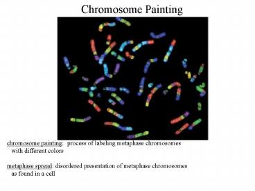
Chromosome Painting
chromosome painting process of labeling
metaphase chromosomes with different colors
metaphase spread disordered presentation of
metaphase chromosomes as found in a cell
2
Human Karyotype
karyotype ordered presentation of metaphase
chromosomes arranged systematically in pairs
humans have 23 pairs of chromosomes 22
'identical' pairs X and Y differ
Giemsa stain cytological stain commonly used to
show transverse bands (G-bands) on
chromosomes-- each chromosome has a specific
number and spacing of bands
3
Mouse Karyotype
mice have 20 chromosomes X and Y are closer in
size chromosome number does not relate to
complexity
4
Karyotypes
Chromosomes have 2 arms on each side of the
centromere short arm or p arm (petit). long
arm is q (not-p) bands farther from the
centromere have higher numbers some bands can
have yet smaller subdivisions numbered
consecutively following the main band number ie.
1p17.2 chromosome 1, short arm, band 17,
sub-band 2 10q22 represents chromosome 10,
long arm, band 22 with the human genome
sequence, each chromosome has a known number,
density, and sequence of genes
5
Karyotypes
centromeres of chromosomes can be found in
characteristic positions on a chromosome
chromosomes without a centromere are
unstable metacentric centromere is roughly in
the middle of a chromosome submetacentric
centromere is somewhat off center but not too
far acrocentric centromere is dramatically
closer to one end than the other
metacentric
submetacentric
acrocentric
6
Karyotypes
acentric chromosome lacking a centromere very
defective and unstable-- chromosome doesnt
separate properly during metaphase because
the spindle fibers cannot attach dicentric
chromosome with 2 centromeres also unstable--
not separated consistently and can often break
if near each other, may act as one and behave
mostly normally cancer cells often have
an abnormal number of chromosomes
7
Cell Cycle
4 stages of cell cycle G1, S, G2, M G1 cell
resting no DNA synthesis S DNA synthesis and
chromosome duplication G2 gap 2, double normal
DNA level M mitosis chromosomes visible, cell
actively dividing (.5-2 hrs)
Interphase
time for various stages varies by cell
type cells growing in culture double 18-24 hours
8
Cell Cycle
cell cycle is under the genetic control of
cyclin-CDK complexes CDK stands for cyclin
dependent protein kinase protein kinase enzyme
which adds a phosphate group to specific
hydroxyls on a target protein 3 amino acid
targets-- serine and threonine, also
tyrosine phosphorylation is the most common
regulatory mechanism in cells-- different
locations can have different effects on the
enzymes phosphatase removes phosphates from
proteins opposite of kinases
9
Cell Cycle
Cyclin family of proteins whose abundance
regulatesvand varies with the cell
cycle cyclin-CDK complexes phosphorylate
transcription factors that transcribe the
next cyclin of the cell cycle as well as other
genes previous cyclin mRNA and protein are
degraded by the ubiquitin proteosome pathway
10
Cell Cycle
eukaryotic cells have multiple different cyclins
and CDK complexes that form different binding
pairs depending upon the cell cycle CDK
complexes well conserved in evolution-- human
gene substitutes fine for its yeast ortholog
11
Cell Cycle Regulation
checkpoint place in the cell cycle where
process can be halted if all required steps have
not been completed start G1 checkpoint-- cell
prepares to synthesize DNA origins of
replication slowly form pre-replication
complexes Rb protein retinoblastoma protein
holds cell in G1 by inhibiting the
transcription factor E2F often mutated in
cancers-- anti-oncogene (prevents cancer
formation) G1/S transition last checkpoint
before the cell must replicate is controlled
by transcription factor E2F DNA damage
checkpoints can interrupt the cell cycle at
multiple points ie. single strand breaks blocks
G1/S, double strand breaks block G2/M centrosome
division checkpoint delays mitosis if not
completed
12
Stages of Mitosis
mitosis cell division that maintains the number
of chromosomes type of division carried out
by most cells
1) Prophase condensation of chromosomes with
paired chromatids chromatids held together at
the centromere
During prophase, chromosomes become shorter and
thicker end of prophase is marked by the loss
of the nuclear membrane
13
Stages of Mitosis
2) Metaphase begins with formation of the
mitotic spindle originating from 2
centrosomes mitotic spindle bundles of
microtubules extending from the centrosome to
centromere of chromosomes kinetochore structure
at the centromere which binds the
spindle kinetochores of each chromosome line
up along a plane equidistant from centrosomes
plane is called the metaphase plate spindle
checkpoint monitors spindle and kinetochore
attachment
14
Stages of Mitosis
3) Anaphase chromatids separate and are pulled
by the kinetochore toward opposite
centrosomes 4) Telophase nuclear
envelope reforms and the mitotic
spindle disappears Chromosomes decondense and a
cleavage furrow separates the cytoplasm in half
15
Meiosis
during reproduction, though, Mendel noted that
gametes only get 1 determinant... if
'determinant' is replaced by 'chromosome', then
gametes should get 1/2 the normal number of
chromosomes Meiosis process of reducing in
half the number of each chromosome only occurs
during the formation of gametes
adult
gametes
meiosis occurs immediately after a 'normal' cell
divison-- there is no opportunity for a second
round of DNA synthesis in S phase
16
Meiosis
meiocyte cells which can undergo meiosis
(primary spermatocytes and oocytes) in
females, only 1 of 4 haploid daughter cells
survive in plants, haploid daughters form
spores, which can then replicate by mitosis to
form a haploid gametophyte organism gametophytes
form gametes by mitosis fusion of haploid
gametes creates diploid zygote forming
sporophyte meiosis takes much longer than
mitosis-- generally days to weeks
17
Meiosis
In cells undergoing meiosis, the first prophase
division occurs in a somewhat different way than
during mitosis initial event of chromosomes
lining up next to each other allows further
cytological distinctions to be made individual
steps are known as leptotene, zygotene,
pachytene, diplotene, diakinesis all 5 steps
occur during prophase I-- steps in
condensation these subdivisions are important
because it allows the exchange of genetic
information between strands by recombination reco
mbination allows individual gametes to combine
traits inherited from the father and the mother
18
Stages of Meiosis
1) Leptotene initial phase of
condensation appears as thin threads with
irregular dense granules (chromomeres) chromomere
s have a characteristic size and number for a
given chromosome 2) Zygotene lateral pairing
of homologous chromosomes (synapsis) bivalent
synapsed homologous chromosome
19
Stages of Meiosis
3) Pachytene further chromosome condensation tet
rad pairing of 4 chromosomes crossing over
begins crossing over event of genetic exchange
between chromosomes 4) Diplotene chromosomes
begin to separate crossing over visible chiasma
cross connection of two chromosomes caused by
breakage and rejoining between chromatids
20
Stages of Meiosis
5) Diakinesis homologous chromosomes not
connected by chiasmata repel each
other chromosomes maximally condensed chiasmata
persist until first anaphase ie. chromatids
still attached! last stage- nuclear membrane
breakdown
21
Results of Meiosis
grandparents
grandparents gametes no recombination
parents
parents gametes after recombination
child is a mixture of both parents and
grandparents recombination is random-- different
children will have different parts of each
chromosome to mix and match traits

