Time scales and molecular motions - PowerPoint PPT Presentation
1 / 54
Title:
Time scales and molecular motions
Description:
Time scales and molecular motions. Atomic fluctuations, vibrations. 10-15 to 10 ... Molecular rotation, reorientation Relaxation, linewidths, correlation times ... – PowerPoint PPT presentation
Number of Views:63
Avg rating:3.0/5.0
Title: Time scales and molecular motions
1
Dynamics and Relaxation
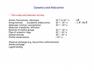
- Time scales and molecular motions
- Atomic fluctuations, vibrations. 10-15 to
10-12 s lt1Å - Group motions. (covalently linked units)
10-12 10-3 s lt 1 Å 50 Å - Molecular rotation, reorientation 10-12 10-9 s
- Molecular translation, diffusion
- Rotation of methyl groups. 10-12 10-9 s
- Flips of aromatic rings. 10-9 10-6 s
- Domain motions. 10-8 10-3 s
- Proline isomerization. gt 10-3 s
- Chemical exchange (e.g. two protein
conformations) - Amide exchange
- Ligand binding
2
Dynamics and Relaxation
- Time scales and molecular motions
- Atomic fluctuations, vibrations. Influences
bond length measurements - Group motions. (covalently linked units)
- Molecular rotation, reorientation Relaxation,
linewidths, correlation times - Molecular translation, diffusion DOSY NMR
- Rotation of methyl groups. 2H NMR
- Flips of aromatic rings. 2H NMR
- Domain motions. 2H NMR
- Chemical exchange, proline isomerization Chemical
shifts - Amide exchange 15N-1H HSQC
- Ligand binding Transferred NOE measurements
3
Dynamics and Relaxation
- Molecular Rotation
- T1 and T2 relaxation times
- Chemical exhange - kinetics
- Amide exchange, chemical shift changes
- Molecular Translation-Diffusion
- DOSY - Diffusion ordered NMR
4
Long
T1 and T2 at short correlation times
T1
T1 or T2 relaxation time
T1 minimum
T2
Short
Fast motion Short tc
Slow motion Long tc
Correlation time
optimal frequency for T1 relaxation (MHz
frequencies)
5
Concept 6 When the B1 field is turned off, the
net magnetization relaxes back to the Z axis with
the time constant T1
T1 is the longitudinal relaxation time constant
which results from spin-lattice relaxation
6
(No Transcript)
7
(No Transcript)
8
Measure T1 with an inversion recovery pulse
sequence 180 - t - 90 - measure intensity
acquire spectrum
acquire spectrum
9
180
Inversion Recovery
10
Inversion Recovery - Measure NMR Intensity as a
function of the delay time t
0
t
11
Inversion Recovery - Measure NMR Intensity as a
function of the delay time t and fit to an
exponential function
0
t
Mz
Mz Mo (1- 2e -t/T1 )
0
t
12
T2 - spin-spin or transverse relaxation
13
Recall first lecture. One spin precessing in the
x-y plane will induce an oscillating current
Current amplitude
time
14
Recall first lecture. Fourier transform of an
infinite sine wave is a delta function, ie. a
sharp line with a single frequency
Current amplitude
frequency
time
15
Now imagine you have many spins with
different orientations in the x-y plane and all
precessing at the same frequency. The net
current is zero and therefore there is no
signal.
Current amplitude
time
time
16
Immediately after all of spins are put into the
X-Y plane, they are in phase.
Current amplitude
time
17
The time it takes for the Individual spins to
dephase in the x-y plane is the T2 relaxation
time.
Current amplitude
time
18
Current amplitude
frequency
time
Current amplitude
frequency
time
Fourier transform of a decaying sine wave gives a
broad line in the frequency domain.
19
Current amplitude
frequency
time
The faster the dephasing, the faster the decay of
the time domain signal, the broader the line.
Line widths are related to T2 relaxation. LW
1/ T2 T2 is always faster (shorter) than or
equal to T1
20
H
H
H
H
H
C
H
H
H
H
C
H
C
H
C
H
C
N
C
C
C
N
C
H
N
C
C
H
H
N
C
H
H
H
H
H
1H spectrum
15N
2D HSQC yields one resonance for each amide N-H
1H
21
Line width at half maximum
15N spectrum
15N
1H
22
T2 relaxation, line width and correlation times
hw viscosity of the solvent
r3H hydrated radius
25
20
Dn FWHM (Hz)
15
10
5
0
2
6
8
10
12
14
4
tc (ns)
See Cavanagh et al. Protein NMR spectroscopy,
pages 16-19.
23
Relaxation, line width and correlation times
hw viscosity of the solvent
r3H hydrated radius
For ubiquitin, 76 residues Mw 8565 1H line
width 6 Hz tc 4.0 ns rH 17 Å Correct
for ubiquitin monomer. What would the line width
be for a tetramer?
24
mobile, flexible chain has narrower line widths
than globular protein
15N
1H
25
Mobility is also expressed in T1 relaxation times.
t 10 us
t 100 us
t 1000 us
t 5000 us
26
Secondary Structure
Sequence MALRRVETTYGDAWCSTQNLIVWRSTERLN
daN 3JHNa gt 7 Hz 1/T1 or 1/T2
loop
sheet
sheet
27
Dynamics and Relaxation
- Molecular Rotation
- T1 and T2 relaxation times
- Chemical exhange - kinetics
- Amide exchange, chemical shift changes
- Molecular Translation-Diffusion
- DOSY - Diffusion ordered NMR
28
Amide Exchange
15N-1H HSQC
Add D20 and collect time series of spectra
29
D20
mobile, flexible chain is more exposed to
solvent and will exchange faster
D20
D20
D20
15N
1H
30
Chemical Exchange
Slow exchange - two distinct resonances
31
Chemical Exchange
32
mobile, flexible chain transiently forms
helix in the limit of slow exchange, you will
observe two distinct resonances
15N
What is slow?
1H
33
Chemical Exchange
Fast exchange - one sharp average resonance
temp
Intermediate exchange - one broad resonance
Slow exchange - two distinct resonances
34
mobile, flexible chain forms helix upon
phosphorylation You can measure the kinetics by
NMR
P
P04-2
1H
1H
35
Kinetics
O
O
H
N
N
OH
time
36
Dynamics and Relaxation
- Molecular Rotation
- T1 and T2 relaxation times
- Chemical exhange - kinetics
- Amide exchange, chemical shift changes
- Molecular Translation-Diffusion
- DOSY - Diffusion ordered NMR
37
NMR magnet.
B
1
Bo
NMR probe
e-
e-
38
NMR magnet.
B
1
Bo
sample
NMR probe
e-
e-
39
NMR magnet.
B
1
Bo
NMR probe
sample
With shimming
Without shimming
40
Gradient pulses are important in diffusion and MRI
B
1
Bo
sample
e-
Gradient pulses can create gradient fields
NMR probe
e-
41
Molecular Diffusion
Hahn echo in absense of gradient pulses
180y
90x
reverse gradient
gradient pulse
t2
t1
If there is no diffusion, a second gradient
pulse, will result in full Hahn echo.
With gradient pulse, magnetization evolves at
different frequencies (it is labeled depending
on its location).
DOSY diffusion ordered nmr spectroscopy (see
Prog. Nucl. Mag. Reson. Spec. 34203)
42
What is Magnetic Resonance Imaging?
- Imagine each slice is divided into separate
voxels
43
What is Magnetic Resonance Imaging?
- Inside of each voxel is water.
44
N
S
- The nucleus of the H in H2O acts like a small bar
magnet
45
What is Magnetic Resonance Imaging?
S
N
- If you place water in a large magnet, the small
bar magnets will align.
46
What is Magnetic Resonance Imaging?
91.2 MHz
S
N
N
S
- You can flip the bar magnets with a pulse of
radiowaves.
47
What is Magnetic Resonance Imaging?
When the bar magnets return, they emit a
radio signal
91.2 MHz
48
What is Magnetic Resonance Imaging?
fast
91.2 MHz
slow
Receiver picks up emitted signals
- Different tissues will return to equilibrium at
- different times.
49
What is Magnetic Resonance Imaging?
91.2 MHz
receiver
- The problem is that you cant tell WHERE the
different signals come from.
50
What is Magnetic Resonance Imaging?
91.2 MHz
102.4 MHz
surface
deep
receiver
- Lauterburs genius was to put a magnetic field
gradient across the sample.
51
What is Magnetic Resonance Imaging?
110.4 MHz
receiver
86.2 MHz
- Gradients can be applied in 3 dimensions.
52
What is Magnetic Resonance Imaging?
110.4 MHz
receiver
91.2 MHz
- This allows one to tell exactly from which voxel
each signal is coming from.
53
What is Magnetic Resonance Imaging?
110.4 MHz
receiver
91.2 MHz
- This allows one to tell exactly from which voxel
each signal is coming from.
54
What is Magnetic Resonance Imaging?
- Each voxel is then color-coded with how fast the
water returns to equilibrium


