hvs PowerPoint PPT Presentation
1 / 44
Title: hvs
1
(No Transcript)
2
Computer Graphics- The Human Visual System -
- Marcus Magnor
3
Overview
- Today
- The Human Visual System
- The eye
- Early vision
- High-level analysis
- Color perception
4
Light
- Electromagnetic radiation
- Visible spectrum 400 to 700 nm
5
Radiation Law
- Physical model for light
- Wave/particle-dualism
- Electromagnetic radiation wave model
- Photons Ephh? particle model ray
optics - Plenoptic function
- L L(x, ?, t, ?, ?), 5 dimensional,
6
Photometry
- Equivalent units to radiometry
- Weight with luminous efficiency function
V(?)(luminous efficiency function) - Spectral or total units
- Distinction in English simple
- rad radiometric unit
- lum photometric unit
7
Radiometric Units
8
Photometric Units
- With luminous efficiency function weighted units
9
Illumination samples
- Typical illumination intensities
10
Human Visual System
- Physical structure well established
- Perceptual behaviour is a complex process
11
HVS - Relationships
Psychophysics
Perception
Stimulus
Neural response
Physiology
12
Perception and Eye
13
Retina
14
Eye
- Eye
- Fovea Ø 1-2 visual degrees
- 6-7 Mio. cones, circa 0.4 arc seconds sized
- No rods
- Three different cone types L, M, S
- Linked directly with nerves
- Resolution 10 arc minutes (S, blue), 0.5 arc
minutes (L, M) - Adaptation of light intensity only through cones
- Periphery
- 75-150 Mio. rods, night vision, S/W
- Response to stimulation of approx. 5 photons/sec.
(_at_ 500 nm) - Many thousands of cells are linked with nerves
- Bad resolution
- Good flickering sensitivity
15
Visual Acuity
Resolution in line-pairs/arc minute
Receptor density
16
Resolution of the Eye
- Resolution-experiments
- Line pairs 50-60/degree ? resolution .5 arc
minutes - Line offset 5 arc seconds 1/6 !! (hyperacuity)
- Eye micro-tremor 60-100 Hz, 5 ?m (2-3
photoreceptor spacings) - Super-resolution
- 19 display at 60 cm 18.000 x 18.000 (3000 x
3000) Pixel - Eye fixates itself
- Automatic gaze tracking
- Overall high resolution
- Visual acuity increased by
- Brighter objects
- High contrast
17
Luminance Contrast Sensitivity
Campbell-Robson contrast sensitivity chart
18
Contrast Sensitivity
- Sensitivity 1 / threshold contrast
- Maximum acuity 5 cycles/degree (0.2 )
- Decrease toward low frequencies lateral
inhibition - Decrease toward high frequencies sampling rate
(Poisson disk) - Upper limit 60 cycles/degree
- Medical diagnosis
- Glaucoma (affects peripheral vision low
frequencies) - Multiple sclerosis (affects optical nerve
notches in contrast sensitivity)
www.psychology.psych.ndsu.nodak.edu
19
Color Contrast Sensitivity
- Color vs. luminance vision system
- Higher sensitivity at lower frequencies
- High frequencies less visible
- Image compression
20
Threshold Sensitivity Function
- Weber-Fechner Law
- Perceived brightness log (radiant intensity)
- EKc log Iv
- Perceivable intensity difference
- 10 cd vs. 12 cd DL2cd
- 20 cd vs. 24 cd DL4cd
- 30 cd vs. 36 cd DL6cd
21
Weber-Fechner Examples
22
Mach Bands
- Overshooting along edges
- Extra-bright rims on bright sides
- Extra-dark rims on dark sides
- Lateral Inhibition
23
Lateral Inhibition
- Pre-processing step within retina
- Surrounding brightness level weighted negatively
- A bright stimulus, maximal bright inhibition
- B bright stimulus, partial bright inhibition gt
stronger response - C dark stimulus, partial dark inhibition gt
weaker response - D dark stimulus, maximal dark inhibition
- High-pass filter
- Enhances contrast along edges
- Difference-of-Gaussians (DOG) function
24
Lateral Inhibition Hermann Grid
- Dark dots at crossings
- Explanation
- Crossings (A)
- More surround stimulation (more bright area)
- More inhibition
- Weaker response
- Streets (B)
- Less surround stimulation
- Less inhibition
- Greater response
- Filtered with DOG function
- Darker at crossings, brighter in streets
- Appears more steady
- What if reversed ?
25
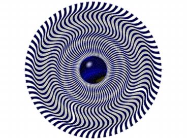
Psychedelic
26
High-level Contrast Processing
27
High-level Contrast Processing
28
Shape Perception
- Depends on surrounding primitives
- Directional emphasis
- Size emphasis
http//www.panoptikum.net/optischetaeuschungen/ind
ex.html
29
Shape Processing Geometrical Clues
http//www.panoptikum.net/optischetaeuschungen/ind
ex.html
- Automatic geometrical interpretation
- 3D perspective
- Implicit scene depth
30
Visual Proofs
http//www.panoptikum.net/optischetaeuschungen/ind
ex.html
31
HVS High-Level Scene Analysis
- Experience
- Expectation
- Local clue consistency
http//www.panoptikum.net/optischetaeuschungen/ind
ex.html
32
Impossible Scenes
- Escher et.al.
- Confuse HVS by presenting contradicting visual
clues
http//www.panoptikum.net/optischetaeuschungen/ind
ex.html
33
Single Image Random Dot Stereograms
34
SIRDS Construction
- Assign arbitrary color to p0 in image plane
- Trace from eyepoints through p0 to object surface
- Trace back from object to corresponding other eye
- Assign color at p0 to intersection points p1L,p1R
with image plane - Trace from eyepoints through p1L,p1R to object
surface - Trace back to eyes
- Assign p0 color to p2L,p2R
- Repeat until image plane is covered
35
Color
- Physics
- Continuous spectral energy distribution
- Human color perception
- Cones in retina
- 3 different cone types
- Spectral mapping to 3 channels
36
Visual Acuity and Color Perception
Mesopic/photopic transition
Photopic vision
Scotopic/mesopic transition
Scotopic vision
37
Color Comparison
- Luminance
- Compare a color source with a gray source
- Luminous Efficiency Function
- Average value from the
- spectral sensitivity ofall receptors
- Photopic day vision (cones)
- Scotopic night vision (rods)
- Mesopic mixed conditions (rods and cones)
Luminous Efficiency Function (V)
38
Color Perception
- Di-chromaticity (dogs, cats)
- Yellow blue-violet
- Green, orange, red indistinguishable
- Tri-chromaticity (humans, monkeys)
- Red, green, blue
- Color-blindness
- Most often men, green color-blindness
www.lam.mus.ca.us/cats/color/
www.colorcube.com/illusions/clrblnd.html
39
Color Mapping
- Spectrum mapping onto perceptual color space
- Infinitely many wavelengths gt 3 color channels
- Cone absorption spectra (S,M,L)
- Overlap of absorption characteristics
- Metamerism
- Same perceived color for different spectral
distributions - Grassmanns law
- Any perceivable color can be represented as a
mixture of three primary colors - Colors add linearly
- From tri-stimulus at every wavelength, total
response can be calculated by integration - But Tri-stimulus response NOT proportional to
absorption spectrum !
40
Standard Color Space CIE-RGB
- Wide range of colors can be mixed from three
monochromatic primary colors 438.1, 546.1, and
700 nm - Colors in the vicinity of 500 nm can only be
matched by subtracting certain amount of r(?) - Inhibitory behavior (gt contrast !)
- Negativecolor values
RGB are called tristimulus values
Color-matching functions for given monochromatic
primary colors
41
Standard Color Space CIE-XYZ
- Standardized imaginary primaries CIE XYZ (1931)
- Non-realizable super-saturated primary colors
- Reproduces all perceivable colors by additive
mixing - Only positive weights
- Y is equivalent to luminance
- Perceivable colors span irregular cone in XYZ
space
42
Chromaticity Diagram
- Normalization
- Projection on the planeof the prime valences
- z 1-x-y
- Chromaticity diagram2D-Plot over x and y
- Points called as color
- locations
- White point (0.3, 0.3)
- Device dependent
- Adaptation of the eye
- Saturation Distanceto the white point
- Complement colors oppositewhite point
The xy chromaticity diagram
White-point line for blackbody radiation
Weißpunkt
43
Wrap-up
- Radiometric vs. photometric units
- Anatomy of the eye
- Rods, cones
- Fovea, blind spot
- Contrast perception
- Weber-Fechner law
- Mach bands, lateral inhibition
- Shape perception
- High-level image analysis
- Color perception
- Tri-stimulus values
- Grassmanns law
- CIE-XYZ standard color space
- Chromaticity diagram
44
(No Transcript)

