Root Fracture - PowerPoint PPT Presentation
1 / 6
Title:
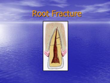
Root Fracture
Description:
Root Fracture Root Fracture Clinical findings: Coronal segment may be mobile/displaced. Percussion positive. Bleeding from the sulcus Radiographic findings ... – PowerPoint PPT presentation
Number of Views:96
Avg rating:3.0/5.0
Title: Root Fracture
1
Root Fracture
2
Root Fracture
- Clinical findings Coronal segment may be
mobile/displaced. Percussion positive. Bleeding
from the sulcus - Radiographic findings horizontal fractures
detected with 90 degree film. Oblique fracture (
often in apical third) require occlusal view or
varying horizontal radiographs. - Treatment Reposition coronal segment promptly.
Confirm reposition radiographically. Splint for 4
weeks. Fractures in cervical third require
stabilization up to 4 months. Monitor pulpal
status for one year. If necrotic endodontic
therapy on the coronal segment to the fracture
line.
3
Root Fracture follow up
- Follow up 4 weeks remove splint, clinical and
radiographic exam. 8 weeks clinical and
radiographic exam. 4 months remove splint on
fractures in the cervical third. Then clinical
and radiographic exam at 6 months, 1 year and 5
years. - Favorable outcome positive pulp test at 3
months. Signs of repair of the fractured
segments. - Negative outcome Symptomatic, negative pulp
test, extrusion of coronal segment, radiolucency
at fracture line. Need for endodontic therapy.
4
Alveolar Fracture
5
Alveolar Fracture
- Clinical findings fracture involves the
alveolar bone and may extend to adjacent bone.
Segment mobility and dislocation with several
teeth moving together. Drastic change in
occlusion. - Radiographic findings Several PA angulations,
occlusal film and panoramic radiograph needed to
determine the course and position of fracture
lines. - Treatment Reposition segment and splint, suture
gingival lacerations. Stabilize segment for 4
weeks
6
Alveolar Fracture follow up
- Follow up 4 weeks remove splint do clinical and
radiographic exam. Follow closely with clinical
and radiographic exam at 8 weeks, 4 months, 6
months, 1 year and 5 years. - Favorable outcome positive pulp tests after 3
months. No signs of PA pathology - Unfavorable outcome Symptomatic negative pulp
test after 3 months. PA lesion or external
inflammatory root resorption. Endodontic therapy
needed.


