
Tissues PowerPoint PPT Presentation
1 / 24
Title: Tissues
1
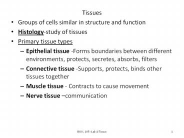
- Tissues
- Groups of cells similar in structure and function
- Histology-study of tissues
- Primary tissue types
- Epithelial tissue -Forms boundaries between
different environments, protects, secretes,
absorbs, filters - Connective tissue -Supports, protects, binds
other tissues together - Muscle tissue - Contracts to cause movement
- Nerve tissue communication
2
- Functions of epithelial tissue
- Protection-covers all exposed body surfaces and
lines all body cavities - Absorption
- Filtration
- Excretion
- Secretionforms endocrine and exocrine glands
- Sensory reception
3
- Characteristics of Epithelial Tissue
- Cells have polarity an apical surface (upper or
free surface) and basal surface (lower or
attached surface) - Cells near apical surface differ from those at
basal surface - Are composed of closely packed cells which are
connected to each other and evenly spaced - Continuous sheets held together by tight
junctions and desmosomes (anchoring) junctions - Supported by connective tissue
- Avascular but innervated-depends on diffusion of
nutrients from basement membrane - High rate of regeneration
4
- Classification of Epithelial tissue
- Number of tissue layers
- Simple one layer of cells
- typically found where absorption and filtration
occur and a thin epithelial barrier is desirable - Stratified many layers of cells
- common in high-abrasion areas where protection is
important i.e. lining of the mouth, skin surface - May have specialized names
- Endotheliuminnermost lining
- Provides a slippery lining
- Mesotheliummiddle lining
- Found in serous membranes lining ventral body
cavity and covering the organs
5
- Classification of Epithelial Tissues
- Type of cell
- Simple
- Squamous-thin, flat cell
- Cuboidal-cube shaped cell
- Columnar tall, rectangular shaped cell
- Stratified
- named according to number of cell layers
6
- Pseudostratified columnar epithelium appears
stratified, typically with nuclei located in at
least two more-or-less distinct levels. But
every cell actually rests on the basement
membrane, so epithelium is technically "simple",
despite appearances.
http//www.siumed.edu/dking2/intro/epith.htm
7
- Glandular Epithelial Tissue
- A gland is one or more cells that makes and
secretes a product called a secretion - Classified by
- Site of product releaseinto duct (exocrine) or
directly into bloodstream (endocrine) - or number of cells forming glandunicellular
(e.g., goblet cells) or multicellular
8
- Endocrine Glands
- Ductless glands
- Secrete hormones that travel through lymph or
blood to target organs - Exocrine Glands
- More numerous than endocrine glands
- Secrete products into ducts
- Secretions released onto body surfaces (skin) or
into body cavities - Examples include mucous, sweat, oil, and salivary
glands
9
- Connective Tissue
- Most abundant and widely distributed tissue type
- Four classes
- Connective tissue proper
- Cartilage
- Bone tissue
- Blood
- Major Functions of Connective Tissue
- Binding and support
- Protection
- Insulation
- Transportation (blood)
10
- Connective Tissue Characteristics
- Connective tissues have
- Varying degrees of vascularity
- Living cells separated by nonliving extracellular
matrix (ground substance and fibers) cells are
scattered and not attached to each other - Allows tissue to be stronger
11
- Structural Elements of Connective Tissue
- Ground substance or Matrix
- Medium through which solutes diffuse between
blood capillaries and cells - Components
- Interstitial fluid
- Adhesion proteins (glue)
- Proteoglycans
- Protein core large polysaccharides
- Trap water in varying amounts, affecting
viscosity of ground substance
12
- Structure Elements of Connective Tissue
- Three types of fibers
- Collagen (white fibers)
- Strongest and most abundant type
- Provides strength
- Elastic
- Networks of long, thin, elastin fibers that allow
for stretch (elasticity) - Reticular
- Short, fine, highly branched collagenous fibers
- Cells
- Mitotically active and secretory cells blasts
- Mature cells cytes
13
- Areolar Connective Tissue
- Gel-like matrix with fibroblasts, macrophages,
mast cells, and some white blood cells. - Function Wraps and cushions organs macrophages
phagocytize bacteria plays important role in - inflammation
- Location Widely distributed under epithelial
tissues e.g.,packages organs surrounds
capillaries. - Adipose Connective Tissue
- Description Matrix sparse nucleus of closely
packed adipocytes pushed to the side by large fat
droplet. - Function reserve fuel insulates against heat
loss supports and protects organs. - Location Under skin in hypodermis around
kidneys and eyeballs within abdomen in breasts
14
- Reticular Connective Tissue
- Description Network of reticular fibers in a
loose ground substance reticular cells lie on
network. - Function Fibers form a soft internal skeleton
that supports other cell types including white
blood cells, mast cells, and macrophages. - Location lymph nodes, bone marrow, and spleen
- Dense, Regular Connective Tissue
- Description parallel collagen fibers with few
elastic fibers major cell type is fibroblast. - Function Attaches muscles to bones or to
muscles attaches bones to bones withstands
great tensile stress when pulling force is
applied in same direction as fibers. - Location Tendons, most ligaments, aponeuroses.
15
- Dense Irregular Connective Tissue
- Description irregularly arranged collagen
fibers some elastic fibers major cell type is
fibroblast. - Function Able to withstand tension exerted in
many directions provides structural strength. - Location Fibrous capsules of organs and joints
dermis of the skin submucosa of digestive tract.
16
- Cartilaginous Connective Tissue
- Three types of cartilage
- Hyaline cartilage
- Elastic cartilage
- Fibrocartilage
- Hyaline Cartilage
- Description firm matrix of collagen fibers
chondroblasts produce the matrix when mature,
(chondrocytes) lie in lacunae. - Function Supports and reinforces has cushioning
properties resists compressive stress. - Location embryonic skeleton covers ends of long
bones forms costal cartilages of ribs
cartilages of nose, trachea, and larynx.
17
- Elastic Cartilage
- Description Similar to hyaline cartilage, but
more elastic fibers in matrix. - Function Maintains structure shape while
allowing - great flexibility.
- Location external ear (pinna) epiglottis.
- Fibrocartilage
- Description Matrix similar to but less firm than
hyaline cartilage thick collagen fibers
predominate. - Function Tensile strength with the ability to
absorb - compressive shock.
- Location Intervertebral discs pubic symphysis
discs of knee joint.
18
- Osseous Tissue-Bone
- Description Hard, calcified matrix containing
many collagen fibers osteocytes lie in lacunae.
Very well vascularized. - Function supports and protects provides levers
for muscles stores calcium and other minerals
and fat marrow inside bones is site for blood
cell formation (hematopoiesis). - Location Bones
- Blood
- Description Red and white blood cells in a fluid
matrix - (plasma).
- Function Transport of respiratory gases,
nutrients, wastes, and other substances - Location Contained within blood vessels.
19
- Muscle Tissue
- Skeletal Muscle
- Description Long, cylindrical, multinucleate
cells striated - Function Voluntary movement locomotion
manipulation of environment facial expression
voluntarily controlled - Location skeletal muscles attached to bones or
to skin. - Cardiac Muscle
- Description Branching, striated, uninucleate
cells that connect at specialized
junctions-intercalated discs - Function As it contracts, it propels blood into
the - circulation involuntary control.
- Location walls of heart
20
- Smooth Muscle
- Description Spindle-shaped cells with central
nuclei no - striations cells arranged closely to form
sheets. - Function Propels substances or objects
(foodstuffs, urine, - a baby) along internal passage-ways involuntary
control. - Location walls of hollow organs.
- Nervous Tissue
- Description Neurons are branching cells cell
processes - may be quite long extend from nucleus-containing
cell body contributing to nervous tissue are
nonirritable supporting cells - Function Transmit electrical signals from
sensory receptors to effectors (muscles and
glands) which control their activity - Location Brain, spinal cord, and nerves.
21
- Epithelial Membranes
- covering and lining tissue composed of an
epithelial layer bound to an underlying
connective tissue - Cutaneous membrane (skin)
- Consists of keratinized stratified squamous
epithelium (epidermis) attached to a layer of
dense irregular connective tissue (dermis)
22
- Epithelial Membranes
- Mucous membranes
- Line body cavities open to exterior (e.g.,
digestive and respiratory tracts) - Are moist
- Directly underneath it is loose connective tissue
called the lamina propria
23
- Epithelial Membranes
- Serous Membranes
- Moist membranes in closed ventral body cavity
- Made of simple, squamous epithelium resting on
areolar connective tissue - Secretes thin, clear serous liquid that
lubricates facing surfaces of parietal and
visceral layers so there is no friction as they
move - Parietal serosa line internal body walls
- Visceral serosa cover internal organs
24
- LAB TO DO
- 1. State the four basic tissue categories and the
different tissues in each category. - 2. ID microscope slides and models of each type
of tissue covered in lab and give examples of
where it is found and its function. - Epithelial simple squamous, simple cuboidal,
simple columnar - pseudostratified (ciliated) columnar
- stratified squamous
- Connective Areolar,
- adipose, hyaline cartilage
- dense regular bone
Recommended
