Tissues - PowerPoint PPT Presentation
1 / 49
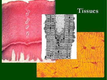
Title: Tissues
1
Tissues
2
Tissues
- Cells work together in functionally related
groups called tissues - How is this done?
- Attachments
- communication
- Types of tissues
- Epithelial lining and covering
- Connective support
- Muscle movement
- Nervous control
3
Epithelial Tissue General Characteristics
Functions
- Covers a body surface or lines a body cavity
- Forms most glands
- Functions of epithelium
- Protection
- Absorption, secretion, and diffusion
- Filtration
- Forms slippery surfaces (mucus secretion)
4
Special Characteristics of Epithelia
- Cellularity
- cells are in close contact with each other with
little or no intercellular space between them - Specialized contacts
- may have junctions for both attachment and
communication - Polarity
- epithelial tissues always have an apical and
basal surface - Support by connective tissue
- at the basal surface, both the epithelial tissue
and the connective tissue contribute to the
basement membrane - Avascular
- nutrients must diffuse from basal layer
- Innervated
- Regenerative
- epithelial tissues are highly mitotic
5
Special Characteristics of Epithelia
6
Classifications of Epithelia
- First name of tissue indicates number of layers
- Simple one layer of cells
- Stratified more than one layer of cells
7
Classifications of Epithelia
- Last name of tissue describes shape of cells
- Squamous cells wider than tall (plate or
scale like) - Cuboidal cells are as wide as tall, as in
cubes - Columnar cells are taller than they are wide,
like columns
8
Naming Epithelia
- Naming the epithelia includes both the layers
(first) and the shape of the cells (second) - i.e. stratified cuboidal epithelium
- The name may also include any accessory
structures - Goblet cells
- Cilia
- Keratin
- Special epithelial tissues (dont follow naming
convention) - Psuedostratified
- Transitional
9
Simple Squamous Epithelium
- Description
- single layer of flat cells with disc-shaped
nuclei - Special types
- Endothelium (inner covering)
- slick lining of hollow organs
- Mesothelium (middle covering)
- Lines peritoneal, pleural, and pericardial
cavities - Covers visceral organs of those cavities
10
Simple Squamous Epithelium
- Function
- Passage of materials by passive diffusion and
filtration - Secretes lubricating substances in serous
membranes - Location
- Renal corpuscles (kidneys)
- Alveoli of lungs
- Lining of heart, blood and lymphatic vessels
- Lining of ventral body cavity (serosae/serous
memb.)
11
Simple Squamous Epithelium
If its from a mesothelial lining
12
Simple Cuboidal Epithelium
- Description
- single layer of cube-like cells with large,
spherical central nuclei - Function
- secretion and absorption
- Location
- kidney tubules, secretory portions of small
glands, ovary surface
13
Simple Cuboidal Epithelium
14
Simple Columnar Epithelium
- Description
- single layer of column-shaped (rectangular) cells
with oval nuclei - Some bear cilia at their apical surface
- May contain goblet cells
- Function
- Absorption secretion of mucus, enzymes, and
other substances - Ciliated type propels mucus or reproductive cells
by ciliary action
15
Simple Columnar Epithelium
- Location
- Non-ciliated form
- Lines digestive tract, gallbladder, ducts of some
glands - Ciliated form
- Lines small bronchi, uterine tubes, and uterus
16
Pseudostratified Columnar Epithelium
- Description
- All cells originate at basement membrane
- Only tall cells reach the apical surface
- May contain goblet cells and bear cilia
- Nuclei lie at varying heights within cells
- Gives false impression of stratification
- Function
- secretion of mucus propulsion of mucus by cilia
17
Pseudostratified Columnar Epithelium
- Locations
- Non-ciliated type
- Ducts of male reproductive tubes
- Ducts of large glands
- Ciliated variety
- Lines trachea and most of upper respiratory tract
18
Stratified Epithelia
- Contain two or more layers of cells
- Regenerate from below
- Major role is protection
- Are named according to the shape of cells at
apical layer
19
Stratified Squamous Epithelium
- Description
- Many layers of cells squamous in shape
- Deeper layers of cells appear cuboidal or
columnar - Thickest epithelial tissue adapted for
protection
20
Stratified Squamous Epithelium
- Specific types
- Keratinized contain the protective protein
keratin - Surface cells are dead and full of keratin
- Non-keratinized forms moist lining of body
openings - Function
- Protects underlying tissues in areas subject to
abrasion - Location
- Keratinized forms epidermis
- Non-keratinized forms lining of esophagus,
mouth, and vagina
21
Stratified Squamous Epithelium
Non-keratinized vs. Keratinized
22
Transitional Epithelium
- Description
- Basal cells usually cuboidal or columnar
- Superficial cells dome-shaped or squamous
- Function
- stretches and permits distension of urinary
bladder - Location
- Lines ureters, urinary bladder and part of
urethra
23
Transitional Epithelium
24
Epithelial Surface Features
- Apical surface features
- Microvilli finger-like extensions of plasma
membrane - Abundant in epithelia of small intestine and
kidney - Maximize surface area across which small
molecules enter or leave - Act as stiff knobs that resist abrasion
25
Epithelial Surface Features
- Apical surface features
- Cilia whip-like, highly motile extensions of
apical surface membranes - Contains a core of nine pairs of microtubules
encircling one middle pair - Axoneme a set of microtubules
- Each pair of microtubules arranged in a doublet
- Microtubules in cilia arranged similarly to
cytoplasmic organelles called centrioles - Movement of cilia in coordinated waves
26
A Cilium
27
Connective Tissue
- Most diverse and abundant tissue
- Main classes
- Connective tissue proper
- Cartilage
- Bone tissue
- Blood
- Components of connective tissue
- Cells (varies according to tissue)
- Matrix
- Fibers (varies according to tissue)
- Ground substance (varies according to tissue)
- dermatin sulfate, hyaluronic acid, keratin
sulfate, chondroitin sulfate - Common embryonic origin mesenchyme
28
Classes of Connective Tissue
29
Connective Tissue Model
- Areolar connective tissue
- Underlies epithelial tissue
- Surrounds small nerves and blood vessels
- Has structures and functions shared by other
connective tissues - Borders all other tissues in the body
- Structures within areolar connective tissue
allow - Support and binding of other tissues
- Holding body fluids
- Defending body against infection
- Storing nutrients as fat
30
Connective Tissue Proper
- Loose Connective Tissue
- Areolar
- Reticular
- Adipose
- Dense Connective Tissue
- Regular
- Irregular
- Elastic
31
Areolar Connective Tissue
- Description
- Gel-like matrix with
- all three fiber types (collagen, reticular,
elastic) for support - Ground substance is made up by glycoproteins also
made and screted by the fibroblasts. - Cells fibroblasts, macrophages, mast cells,
white blood cells - Function
- Wraps and cushions organs
- Holds and conveys tissue fluid
- Important role in inflammation Main battlefield
in fight against infection - Defenders gather at infection sites
- Macrophages
- Plasma cells
- Mast cells
- Neutrophils, lymphocytes, and eosinophils
32
Areolar Connective Tissue
- Location
- Widely distributed under epithelia
- Packages organs
- Surrounds capillaries
33
Adipose Tissue
- Description
- Closely packed adipocytes
- Have nucleus pushed to one side by fat droplet
Function - Provides reserve food fuel
- Insulates against heat loss
- Supports and protects organs
- Location
- Under skin
- Around kidneys
- Behind eyeballs, within abdomen and in breasts
34
Reticular Connective Tissue
- Description network of reticular fibers in
loose ground substance - Function form a soft, internal skeleton
(stroma) supports other cell types - Location lymphoid organs
- Lymph nodes, bone marrow, and spleen
35
Dense Regular Connective Tissue
- Description
- Primarily parallel collagen fibers
- Fibroblasts and some elastic fibers
- Poorly vascularized
- Function
- Attaches muscle to bone
- Attaches bone to bone
- Withstands great stress in one direction
- Location
- Tendons and ligaments
- Aponeuroses
- Fascia around muscles
36
Cartilage
- Characteristics
- Firm, flexible tissue
- Contains no blood vessels or nerves
- Matrix contains up to 80 water
- Cell type chondrocyte
- Types
- Hyaline
- Elastic
- Fibrocartilage
37
Hyaline Cartilage
- Description
- Imperceptible collagen fibers (hyaline glassy)
- Chodroblasts produce matrix
- Chondrocytes lie in lacunae
- Function
- Supports and reinforces
- Resilient cushion
- Resists repetitive stress
38
Hyaline Cartilage
- Location
- Fetal skeleton
- Ends of long bones
- Costal cartilage of ribs
- Cartilages of nose, trachea, and larynx
39
Elastic Cartilage
- Description
- Similar to hyaline cartilage
- More elastic fibers in matrix
- Function
- Maintains shape of structure
- Allows great flexibility
- Location
- Supports external ear
- Epiglottis
40
Fibrocartilage
- Description
- Matrix similar, but less firm than hyaline
cartilage - Thick collagen fibers predominate
- Function
- Tensile strength and ability to absorb
compressive shock - Location
- Intervertebral discs
- Pubic symphysis
- Discs of knee joint
41
Bone Tissue
- Function
- Supports and protects organs
- Provides levers and attachment site for muscles
- Stores calcium and other minerals
- Stores fat
- Marrow is site for blood cell formation
- Location
- Bones
42
Blood Tissue
- Description
- red and white blood cells in a fluid matrix
- Function
- transport of respiratory gases, nutrients, and
wastes - Location
- within blood vessels
- Characteristics
- An atypical connective tissue
- Develops from mesenchyme
- Consists of cells surrounded by nonliving matrix
43
Muscle Tissue
- Types
- Skeletal muscle tissue
- Cardiac muscle tissue
- Smooth muscle tissue
44
Skeletal Muscle Tissue
- Characteristics
- Long, cylindrical cells
- Multinucleate
- Obvious striations
- Function
- Voluntary movement
- Manipulation of environment
- Facial expression
- Location
- Skeletal muscles attached to bones (occasionally
to skin)
45
Cardiac Muscle Tissue
- Function
- Contracts to propel blood into circulatory system
- Characteristics
- Branching cells
- Uninucleate
- Striations
- Intercalated discs
- Location
- Occurs in walls of heart
46
Smooth Muscle Tissue
- Characteristics
- Spindle-shaped cells withcentral nuclei
- Arranged closely to form sheets
- No striations
- Function
- Propels substances along internal passageways
- Involuntary control
- Location
- Mostly walls of hollow organs
47
Nervous Tissue
- Function
- Transmit electrical signals from sensory
receptors to effectors - Location
- Brain, spinal cord, and nerves
- Description
- Main components are brain, spinal cord, and
nerves - Contains two types of cells
- Neurons excitatory cells
- Supporting cells (neuroglial cells)
48
Tissue Response to Injury
- Inflammatory response non-specific, local
response - Limits damage to injury site
- Immune response takes longer to develop and
very specific - Destroys particular microorganisms at site of
infection
49
The Tissues Throughout Life
- At the end of second month of development
- Primary tissue types have appeared
- Major organs are in place
- Adulthood
- Only a few tissues regenerate
- Many tissues still retain populations of stem
cells - With increasing age
- Epithelia thin
- Collagen decreases
- Bones, muscles, and nervous tissue begin to
atrophy - Poor nutrition and poor circulation poor health
of tissues


