Sound - PowerPoint PPT Presentation
1 / 16
Title: Sound
1
Sound
A mechanical wave is a wave which is not capable
of transmitting its energy through a vacuum.
Mechanical waves require a medium in order to
transport their energy from one location to
another. A sound wave is an example of a
mechanical wave. Sound waves are incapable of
traveling through a vacuum. Slinky waves, water
waves, stadium waves, and telephone chord waves
are other examples of mechanical waves each
requires some medium in order to exist. A slinky
wave requires the coils of the slinky a water
wave requires water a stadium wave requires fans
in a stadium and a telephone chord wave requires
a telephone chord.
2
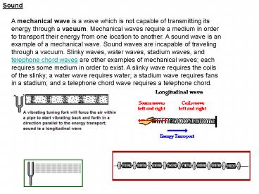
A vibrating tuning fork is capable of creating
such a longitudinal wave. As the tines of the
fork vibrate back and forth, they push on
neighboring air particles. The forward motion of
a tine pushes air molecules horizontally to the
right and the backward retraction of the tine
creates a low pressure area allowing the air
particles to move back to the left. Because of
the longitudinal motion of the air particles,
there are regions in the air where the air
particles are compressed together and other
regions where the air particles are spread apart.
These regions are known as compressions and
rarefactions respectively. The compressions are
regions of high air pressure while the
rarefactions are regions of low air pressure. The
diagram below depicts a sound wave created by a
tuning fork and propagated through the air in an
open tube. The compressions and rarefactions are
labeled.
3
- Describing Sound Waves\
- If sound requires a material medium to travel,
then in sonograghy, the material medium is the
tissue. Before understanding what sonography is
all about, it is important for us to understand
the parameters that describe features of sound. - These include
- Period
- It is the time it takes a wave to vibrate
a single cycle, or time to make a complete
oscillation or time from the start of a cycle to
the start of the next cycle. - It is measured in the unit of time, i.e.
seconds, ms (milliseconds), hrs etc
Period
Period
4
b) Frequency In the wave language, frequency
refers to the number of cycles that occur in one
second. It is reported in the units of per
second (1/second), hertz (Hz). Hertz is another
way to say per second. 1 cycle /second
1 hertz 1,000 cycles/second 1 kHz
1,000,000 cycles/second 1 MHz The higher the
frequency, the shorter the period and vice
versa. The frequency of a sound wave is
determined by the sound source, not by the
medium through which sound travels.
5
More on frequency Infrasonic- sound waves
with frequency of 20Hz or below and cannot be
heard by human ears.
Audible sound waves with frequencies between
20Hz and 20,000 Hz and can
be detected by our human ears. Ultrasonic
sound waves with high frequencies that cannot be
detected by our ears. Has
frequency of 20kHz or higher. Why is frequency
important in diagnostic sonography? It
affects penetration and image quality. c)
Amplitude It is the bigness of a wave. It is
found by subtracting the maximum value and the
average of the undisturbed value of a wave.
Amplitude can take a variety of units, e.g. in
sound, it can take the unit of pressure (Pascal),
or density (g/cm3) or length (cm) etc
6
- Mini-quiz to check your understanding
- Why doesn't sound travel through a vacuum?
- Sound is a waveform in matter, and there is no
matter in a vacuum (a) - Vacuums absorb most of the sound (b)
- It does travel in space, as verified on Star Trek
(c) - 2. How is a sound wave different than a water
wave? - Sound does not travel through water (a)
- Water waves have a wavelength, while sound waves
don't (b) - A water wave moves up-and-down, while sound is a
compression wave (c) - 3. What happens when sound hits a thin membrane?
- It causes the membrane to vibrate (a)
- The membrane reflects most of the sound (b)
- The amplitude of the sound increases (c)
7
d) Power It is the rate of energy transfer
or the rate at which work is performed. It
describes the bigness of a wave. Has a unit
of watts. power amplitude2
(amplitude squared) Example A sonographer
increases the amplitude of a wave by a factor of
3. How has the power
changed? power is proportional to
amplitude squared therefore
32 3x3 9 hence the power is
increased 9-fold e) Intensity It is the
concentration of energy in a sound beam.
Intensity ( w/cm2) power (w) divided by area
(cm2) The intensity of sound changes as sound
propagates through the body and the rate at
which it changes depends on the characteristics
of the medium and the shape of the sound
beam. intensity is proportional to power
i.e. intensity power This implies
that intensity amplitude2
8
f) Wavelength It is the distance or length of
one complete cycle. Its units is in meters (m),
or millimeters (mm), or nanometers (nm), or
micrometers (µm) etc.
Wavelength is the only parameter that is
determined by both the source and the Medium.
9
As long as we remain in one medium, wavelength
and frequency are inversely related. Wavelength
is soft tissue Sound with a frequency of 1
MHz has a wavelength of 1.54 mm The rule for
relationship between frequency and wavelength of
sound in soft tissue Why wavelength
important? It determines image quality.
Shorter wavelength sound usually produces higher
quality images with greater details. g)
Propagation speed Is the distance that
a sound wave travels through a medium in 1
second. Measured in m/s, mm/micosecs,
10
Describing Pulsed Waves (Chapter 4) In
diagnostic ultrasound, continuous wave sound
cannot create anatomic images. Rather, imaging
systems produce short bursts or pulses of a
acoustic energy to create every picture of
anatomy. What is pulsed sound?
A pulse of ultrasound is a collection of cycles
that travel together. A pulse has a
beginning and an end. Although a pulse is made up
of individual cycles, the entire pulse moves
as a single unit. Pulsed ultrasound has two
components transmit, talking or on time
receive, listening, or off time
Off time
On time
On time
11
- In trying to understand pulsed sound, there are
several terms that we need to know - Namely
- Pulse Duration (PD)
- In ultrasound imaging, you use a transducer
to generate the ultrasound pulses - and receive the returning echoes from the
patient. The transducer emits a pulse - with a fixed duration.
- PD is the actual time from the start of
a pulse to the end of the pulse. - It is a single transmit, talking, or on time.
- It is measured in the unit of time, such as
microseconds, milliseconds, or seconds.
12
B) Spatial Pulse Length (SPL) It is the
distance that a pulse occupies in space from the
start to the end of a pulse. It is measured in
the units of distance such as mm, cm
etc.
SPL Number of Cycles (n) x
wavelength (?) What is the difference between
Pulse duration (PD) and Spatial Pulse Length
(SPL)? PD is the time that a pulse is on
and is typically measured in seconds while SPL
is the distance when the pulse is on and is
measured in mm or cm. In diagnostic imaging,
shorter PD and shorter SPL are desirable because
they create more accurate images.
13
C) Listening Time (LT) This is the
time the transducer is receiving signals from
reflectors in the body. By changing the
listening time, sonographers alter the depth of
the image. The shorter the listening
time, the shallow the imaging, while the longer
the listening time, the deeper the
imaging. Depth of view describes the maximum
distance into the body that an ultrasound System
is imaging. PRP is directly related to the depth
of view.
14
D) Pulse Repetition Period (PRP) It is
the time from the start of one pulse to the start
of the next pulse
PRP PD LT Two important components of
PRP are the transit time (PD) and the receive
time (LT). A sonographer cannot change the PD
because it is a characteristic of the
transducer.
15
E) Pulse Repetition Frequency (PRF) It
is the number of pulses that an ultrasound system
transmits into the body each second. With
regard to PRF, the number of cycles are
meaningless. We are just concerned about the
number of pulses created per second. It is
measured in the units of frequency Hz (Hertz) or
per second. It is determined by the maximum
imaging depth of the system. When the system is
imaging shallow, the PRF is higher, while when
the imaging is deep, the PRF is lower. Hence
this is an inverse relationship, ie. PRF and
depth are inversely related. How is the PRP and
PRF related? They are inversely related
Hence PRF 1/PRP or
PRP 1/PRF
16
F) Duty Factor It is the percentage or
fraction of time that the system is transmitting
a pulse. It is a percentage hence
dimensionless (no units). The sonographer
can change the duty factor by altering the image
depth. As image depth increases, trasmit time
(PD) remains constant while listening Time (LT)
is prolonged. As a result, the duty factor
decreases as a system images deeper.


