Types of Microscopy - PowerPoint PPT Presentation
1 / 1
Title:
Types of Microscopy
Description:
Detect reflected light (opaque samples) or transmitted light ... Therefore, it can also be used to etch the sample. Scanning Tunneling Microscopy (STM) ... – PowerPoint PPT presentation
Number of Views:42
Avg rating:3.0/5.0
Title: Types of Microscopy
1
Types of Microscopy
Note this table is intended as a simple guide.
Actual performance and usage may be different in
certain applications.
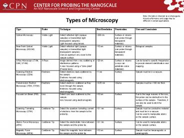
Type Probe Technique Best Resolution Penetration Uses and Constraints
Optical Microscopy Visible Light Detect reflected light (opaque samples) or transmitted light (transparent samples). Light focused using lenses. 200 nm Surface or volume (can probe through transparent materials)
Near-Field Optical Microscopy (NSOM) Visible Light Detect reflected light (opaque samples) or transmitted light (transparent samples). Uses an aperture very close to the sample surface. 10 nm Surface or volume (can probe through transparent materials) Biological samples.
X-Ray Microscopy (TXM, SXM, STXM) X-Rays Image derived from x-ray scattering or interference patterns. X-rays focused using a zone plate (Fresnel lens). 20 nm Surface or volume (x-rays can penetrate some materials) Can be tuned to specific frequencies to provide element identification and mapping.
Scanning Electron Microscopy (SEM) Electrons Detect electrons back-scattered by the sample. Electrons focused using electromagnets. 1 nm Surface Sample must be in a vacuum.
Transmission Electron Microscopy (TEM, STEM) Electrons Detect electrons scattered as they move through the sample. Electrons focused using electromagnets. 0.05 nm Volume Samples must be lt100 nm thick.
Focused Ion Beam (FIB) Ions Detect ions back-scattered by the sample. Ions focused using electromagnets. 10 nm Surface Due to the large masses of the ions, this probe can be destructive to the surface of the sample. Therefore, it can also be used to etch the sample.
Scanning Tunneling Microscopy (STM) Cantilever Tip Detect the quantum tunneling current of electrons from the sample to the probe tip. 0.1 nm Surface Sample must be conductive material and must be in a vacuum. Can be used to manipulate atoms on the sample surface.
Atomic Force Microscopy (AFM) Cantilever Tip Detect the electrostatic force between the sample and the probe tip. 0.1 nm Surface Can be used to manipulate atoms on the sample surface.
Magnetic Force Microscopy (MFM) Cantilever Tip Detect the magnetic force between the sample and the probe tip. 10 nm Surface Sample must be ferromagnetic or paramagnetic.


