Slide sem ttulo PowerPoint PPT Presentation
1 / 1
Title: Slide sem ttulo
1
IN VITRO CHARACTERIZATION OF gE NEGATIVE BOVINE
HERPESVIRUS TYPES 1.1 (BHV-1.1) and 1.2a
(BHV-1.2a) A. C. Franco1,2 F. A. M.
Rijsewijk3 R. Weiblen4 E. F. Flores4 P. M.
Roehe1,5. 1 Centro de Pesquisas Veterinárias
Desidério Finamor, Caixa Postal 47, CEP
92990-000, Eldorado do Sul, RS, Brazil. 2
Universidade Luterana do Brasil, rua Miguel
Tostes, 101, Canoas, RS, Brazil. 3 Institute for
Animal Science and Health, Division of infectious
diseases and food chain quality, (ID-Lelystad),
Postbus 65, 8200 AB Lelystad, The Netherlands. 4
Centro de Ciências Rurais, Universidade Federal
de Santa Maria, CEP 97119-900, Santa Maria, RS,
Brasil. 5 Instituto de Ciências Básicas da Saúde,
Universidade Federal do Rio Grande do Sul, av.
Sarmento Leite, 500, CEP 900150-170, Porto
Alegre, RS, Brazil
Introduction The bovine herpesvirus type 1
(BHV-1) genome encodes several glycoproteins
which are responsible for viral attachment and
entry, cell-to-cell spread or host immune
response evasion (1). The glycoprotein E (gE)
gene is located in the unique short (US) region
of the BHV-1 genome and its role in the in vitro
growth properties of BHV-1.2 viruses have not
yet been addressed. BHV-1.1 gE forms a complex
with gI and is important for cell-to-cell spread
(2,3). In comparison to wild type viruses, gE
negative (gE-) recombinants produce smaller
plaque sizes in vitro, although no alteration of
the growth kinetics or penetration process was
observed for these viruses (2,3). Here, the in
vitro growth characteristics of a brazilian
BHV-1.2a gE- were studied and compared with the
parental virus, as well as with a BHV-1.1 gE-
recombinant, derived from a European BHV-1.1
strain.
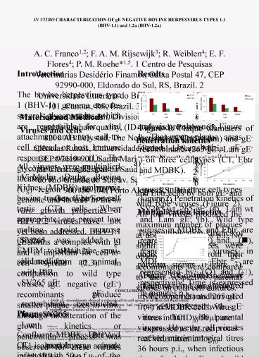
Results Plaque assay The results of the plaque
size analysis are shown in Figure 1. The mean
plaque areas formed by both gE- recombinants were
strikingly reduced when compared to their
respective parental viruses, in all three cell
types (plt0.001).
Material and Methods Viruses and
cells _________________________________________
_____________________ Virus Genotype/Source __
__________________________________________________
___________ SV265 BHV-1.2a isolated from an
animal with IBR SV265 gE- gE- recombinant
constructed from SV265 Lam BHV-1.1 isolated
from an animal with IBR Lam gE- gE-
recombinant constructed from Lam ________________
________________________________________________
Figure 1 Plaque diameters of parental (SV265,
Lam) and gE- recombinants (265 gE-, Lam gE-) on
three cell types (CT, Ebtr and MDBK).
Penetration kinetics Plaque production was first
visualized after 20 minutes of adsorption on both
MDBK and Ebtr cells by both gE- and wild type
viruses (Figure 2). All four viruses produced the
maximum number of plaques at about 120 minutes
p.i. No significant differences were observed
when gE- recombinants were compared to their
respective parental viruses on both cell
cultures.
a)
b)
All viruses were multiplied in Madin Darby Bovine
Kidney (MDBK), embryonic bovine trachea (Ebtr) or
calf testis (CT) cells. When appropriate, one
percent low melting point agarose (Sigma) was
added to EMEM to obtain the semi-solid
medium. Plaque assays Confluent MDBK, Ebtr
and CT monolayers were infected with 50 p.f.u. of
the appropriate virus. After adsorption the
cells were overlaid with semi-solid medium. Four
days post inoculation (p.i.) the cells were
fixated (10 formalin, 1 crystal violet), and
the diameter of at least 50 viral plaques was
measured in each cell type. Penetration
kinetics Penetration was assessed by allowing
approximately 500 p.f.u. of the appropriate virus
to adsorb on either MDBK or Ebtr monolayers. At
different time intervals the inoculum was removed
and the cells were then overlaid with fresh
semi-solid medium and incubated for four days.
Monolayers were then fixed with 10 formalin and
viral plaques were counted at each time interval.
All tests were done in triplicate.
Multiplication kinetics Preformed MDBK cell
monolayers were infected at a m.o.i. of 10.
Adsorption was allowed for 1 hour at 37 C before
the inoculum was removed and extracellular virus
inactivated. The monolayers were incubated for
different intervals (3, 5, 7, 9, 11, 13, 16, 24,
36 and 48 hours p.i.). After the incubation
period, the supernatants were harvested and
assayed for virus. All experiments were performed
in triplicate. Virus titres were calculated
according to the method of Spearmann and Kärber
and expressed as the log10 tissue culture
infectious doses per 50 ?l (TCID50/50?l).
Figure 2 Penetration kinetics of SV265 wt, 265
gE- (a), Lam wt and Lam gE- (b). Wild type
viruses in MDBK and Ebtr are represented by (?)
and (?), respectively. gE- viruses in MDBK and
Ebtr are represented by (?) and (),
respectively. Time is expressed in minutes p.i.
Multiplication kinetics The multiplication
kinetics of both gE- viruses was
undistinguishable from their parental strains
(Figure 3). About 10 times more infectious virus
was detected in the supernatants harvested from
cells infected with gE- viruses than with
parental viruses. However, all viruses reached
maximum viral titres 36 hours p.i., when
infectious titres for both gE- and wild type were
very similar (between 106 and 107 TCID50).
50
Hours post-infection
Figure 3 Growth kinetics of Lam wt (), Lam gE-
(?), SV265 wt (?) and 265 gE- (?) in MDBK cells.
Virus titres in TCID50/50?l, are expressed as the
reciprocal of virus titres in log10.
CONCLUSIONS -Both gE- recombinants showed a
reduced cell-to-cell spread in all three cell
types tested -The absence of gE in both virus
subtypes had no significant effect on the
penetration and multiplication kinetics of the
recombinant viruses
References 1. Spear, P.G. Eisenberg, R.J.
Cohen, G.H. Three classes of cell surface
receptors for alphaherpesvirus entry. Virology,
v. 275 (1), p.1-8. 2000 2 Rebordosa, X. Pinol,
J. Perez-Pons, J.A. Lloberas, J. Naval, J.
Serra-Hartmann, X. Espuna, E. Querol, E.
Glycoprotein E of bovine herpesvirus type 1 is
involved in virus transmission by direct
cell-to-cell spread. Virus Research, v. 45
(1)59-68. 1996 3. Chowdhury, S.I. Ross, C.S.D.
Lee, B.J. Hall, V. Chu, S. Construction and
characterization of a glycoprotein E gene deleted
bovine herpesvirus type 1 (BHV-1) Recombinant
Virus. American Journal of Veterinary Research.,
v. 60, p. 227-232. 1999

