Cervical anatomy - PowerPoint PPT Presentation
1 / 31
Title:
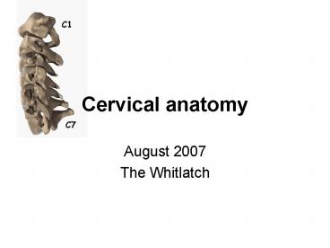
Cervical anatomy
Description:
Superior projects medially and inferior articular facet ... posterior part of the posterior fossa, and posterior tentorium, and posterior falx cerebelli ... – PowerPoint PPT presentation
Number of Views:355
Avg rating:3.0/5.0
Title: Cervical anatomy
1
Cervical anatomy
Mastoid process
C4-5 facet joint
C3 body
C4 body
- August 2007
- The Whitlatch
2
Overview building the cervical spine
- Atlas
- axis
- ligaments
- muscles
- fascia
- the vert
- And how to apply to common cases
3
Atlas
- Ring of bone
- Lateral mass on each side
- Transverse process
- Superior projects medially and inferior articular
facet projects medially to C2 superior artic
facet - 3 cm canal
4
Axis
5
Atlanto-Occipital Joint
- Allows flexion and extension and slight side to
side motion - almost NO rotation
- Stability dependent on ligaments
- ALL, attaches to tubercle on axis, then
small contin to skull - apical ligament
- tectorial membrane (broad ligamentous sheet)
- (continuation of PLL)
- cruciate ligament
- --formed by rostral and caudal longitudinal
bands - Alar ligaments
- arise from dens, connect to medial occipital
condyle - limit rotation of AO joint
- dorsal atlanto-occipital membrane
- (continuation of Ligamentum flavum),
- --remember overlays vert, C1
- C1 and C2 nerves pass dorsally to
occipitocervical and C1/2 joint capsules, NOT
ventral to facets UNLIKE other cervical vertebrae
6
Draw C1, 2 ligaments (coronal view)
A- apical B- alar C-cruciform D-tectoral
7
Draw C1, 2 ligaments (sagittal view)
A- apical B- anterior alantooccipital C-cruciform
D-tectorial membrane (PLL)
8
Movements allowed in the craniocervical
region Range of Joint Motion motion
(degrees) OcciputC1 Combined flexion/extension
25 Lateral bending (unilateral) 5 Axial
rotation (unilateral) 5 C1C2 Combined
flexion/extension 20 Lateral bending
(unilateral) 5 Axial rotation (unilateral) 40
VOLUME 60 NUMBER 1 JANUARY 2007 SUPPLEMENT
9
Surface anatomy of neck
10
Neck triangles
- Anterior triangle
- 4 triangles
- muscular triangle--formed by the midline,
superior belly of the omohyoid, and SCM - carotid triangle--formed by the superior belly of
the omohyoid, SCM, and posterior belly of the
digastric - submental triangle--formed by the anterior belly
of the digastric, hyoid, and midline - submandibular triangle--formed by the mandible,
posterior belly of the digastric, and anterior
belly of the digastric - Posterior triangle
- supraclavicular triangle--formed by the inferior
belly of the omohyoid, clavicle, and SCM - occipital triangle--formed by inferior belly of
the omohyoid, trapezius, and SCM
11
Cervical fascia
- Investing
- surrounds entire neck, splitting to enclose the
SCM and trapezius and parotid glands - Visceral (pretracheal)
- deep to infrahyoid, surrounds visceral space,
including thyroid, trachea and esophagus - attached to hyoid bone and thyroid cartilage
- laterall blends into carotid sheath
- prevertebral
- surrounds vertebral column and muscles
- within prevertebral fascia, anterior, slightly
lateral lie cervical sympathetic plexus, usually
at 1st rib level, C6, and atlantooccipital complex
12
Cervical musculature
- superficial
- platysma, SCM, infrahyoid (sternohyoid,
sternothyroid, omohyoid, and thyrohyoid,
innervated by ansa cervicalis (except
thyrohyoidCN XII)). - infrahyoid group helps swallowing
- deep
- scalene groupanterior, medius, and posterior
- form roof over cupula over lung arise from
transverse process innervated by C4-C8 ventral
rami - longus group--rectus capitis anterior, longus
capitis, longus coli - ventral rami C1-C6, flex head and c spine
13
- deep
- scalene groupanterior, medius, and posterior
- form roof over cupula over lung arise from
transverse process innervated by C4-C8 ventral
rami - longus group--rectus capitis anterior, longus
capitis, longus coli - ventral rami C1-C6, flex head and c spine
14
The Vert
- The paired vertebral arteries arise from the
subclavian aa - They ascend through the transverse processes of
the upper 6 cervical vertebrae - Pass behind the lateral mass of C1 and enter the
dura behind the occipital condyle - Ascend through the foramen magnum and join to
form the basilar artery
15
The Vert, summary of key pts of course
- Ascends through foramen transversaria
- Accompanied by vertebral veins and sympathetic
plexus fibers from cervicothoracic ganglion - Medial to intertransverse muscles
- Lateral bend at atlas
- Curve back on superior surface of atlas
- Between rectus capitis lateralis, superior
articular process of atlas - With the ventral ramus of the first occipital
nerve and curves with it horizontally around
lateral and dorsal aspect of superior articular
process - Transverses articular process, dorsal arch of
atlas, rostal to dorsal ramus of 1st cervical
nerve - Verebral vein originates from plexus of veins
from internal venous plexus and suboccipital
triangle, accompanies vert through foramen
transversaria and exits at (usually) sixth
cervical transverse process
16
The Vert
The extradural part consists of 3 segments
- V1
- origin at subclavian a ? lowest transverse
foramen (usually C6) - V2
- ascending in foramen of the transverse processes
of C6?C1 - V3
- passes medially behind lateral mass of C1 and
across the groove on the upper surface of the
lateral part of the posterior arch of the atlas
then foramen magnum? dura
17
The Vert
V3 passes medially behind lateral mass of C1
and across the grovve on the upper surface of the
lateral part of the posterior arch of the atlas
then foramen magnum? dura
- Partially covered by atlantooccipital membrane,
rectus capitis, and is surrounded by a venous
plexus made up of anastomoses from deep cervical
and epidural vv. - 50 lie in a groove, 50 are surrounded by bone
to some extent, or completely
18
The Vert
Anterior Meningeal Artery
- arises from medial surface of extradural
vertebral artery immediately above the transverse
foramen of C3. - Enters the spinal canal and ascends between the
PLL and dura, at the level of the apex of the
dens, it courses medially to join its mate from
the other side (forming an arch over the tip of
the dens) - Then sends branches to supply the dura in the
region of the clivus, and anterior foramen
magnum, and upper spinal canal - Anastomoses with ascending pharyngeal and dorsal
meningeal aa
19
The Vert
V3 gives off ? branches
- 1) Posterior meningeal from post VA as it courses
around the lateral mass of the atlas- tortuous
course then perforates the dura of the posterior
foramen magnum - Ascends near falx cerebelli and divides near the
torcula into several branches to supply the dura
of the posterior part of the posterior fossa, and
posterior tentorium, and posterior falx cerebelli - 2) Posterior Spinal A (may also arise from
intradural VA or off PICA) courses medially, and
upon reaching the lower medulla, divides into an
ascending and descending branches - Ascending branch through foramen magnum
? restiform body, gracile and cuneat tubercles,
rootlets of XI, and choroid plexus near foramen
of Magandie - Descending branch passes between dorsal
rootlets on posterolateral surface of spinal cord
and supplies the superficial dorsal half of the
spinal cord, and anastomoses with radicular aa.
Also gives off branches to deep cervical mm and
rarely, PICA
20
The Vert intradural segment
- The intradural segment begins at the dural
foramina just inferior to lateral edge of foramen
magnum- the dura here is thicker and forms a
funnel over 4-6mm of the artery, the posterior
spinal aa enter the intradural spinal canal
through this same foramen, and the C1 root leaves
extradurally though this foramen - Once inside the dura, the artery ascents from
lateral ? upper medial surface of medulla - The intradural component is divded into 2
segments lateral medullary and anterior
medullary, - The anterior medullary segment begins at the
preolivary sulcus and passes in front (or
between) rootlets of XII, and crosses the pyramid
to join the contralateral VA to for the basilar A
near the pontomedullary sulcus
Also gives off branches to deep cervical mm and
rarely, PICA
21
On every boards
Anterior-Posterior Anastomoses
- 1) PCom ICA ? PCA
- Fetal origin of PCA PCA arises from PCom
Infundibulum junctional dilation at origin at
ICA - 2) Primitive Trigeminal Artery posterior
cavernous ICA ? Basilar (b/t SCA and AICA) Most
common persistent fetal connection (0.1-0.5) - 3) Persistent Otic Artery petrous ICA through
IAC ? Basilar - Very rare, almost never seen
angiographically - 4) Persistent Hypoglossal Artery cervical ICA
(C1,2)hypoglossal canal ? Basilar Second most
common (0.1), less common than PTA - 5) Proatlantal Intersegmental Artery
Suboccipital cervical ICA ? Vertebral - Runs between arch of C1 and occiput
- Horizontal course along C1 ring
22
Non sequitur
- Non sequitur (IPA n?n 's?kw?t?r) is Latin for
"It does not follow," coming from the dependent
verb sequor. The term may refer to - Non sequitur (logic), logical fallacy
- Non sequitur (rhetoric), a comment which has no
relation to the comment it follows
23
A, a midsagittal section of the cervical spine
configured in lordotic posture (effective
cervical lordosis). A line has been drawn from
the dorsocaudal aspect of the vertebral body of
C2 to the dorsocaudal aspect of the vertebral
body of C7 (solid line). The gray zone is
outlined by the other lines. A midsagittal
section of a cervical spine in kyphosis
(effective cervical kyphosis). B, note that
portions of the vertebral bodies are located
dorsally to the gray zone. C, a midsagittal
section of a straightened cervical spine. Note
that the most dorsal aspects of the vertebral
bodies are located within, but not dorsally to,
the gray zone,
from, Benzel EC Biomechanics of Spine
Stabilization. Rolling Meadows, American
Association of Neurological Surgeons
Publications, 2001 6).
24
Coronal plane tethering (coronal bowstring
effect). A, thenerve roots, or, more commonly,
the dentate ligaments, may tether the spinal cord
in the coronal plane. B, laminectomy may not
relieve the distortion. C, ventral decompression
is a more-commonly considered approach,
(from,Benzel EC Biomechanics of Spine
Stabilization. Rolling Meadows, American
Association of Neurological Surgeons
Publications, 2001 6).
25
Approaches to occipital cervical region
- Dorsal
- Ventral
- Ventral retropharyngeal
- Transoral
- Extended maxillotomy
- Transcondylar
26
Spinous process wiring
Left The Rogers interspinous wiring technique
in a figure-eight pattern. Right Bohlman
triplewire technique. After using the Rogers
technique, two separate wires are threaded
through the holes in upper and lower
spinousprocesses and corticocancellous bone
grafts, which are fastened to the decorticated
bone to promote fusion.
27
Callahan facet wiring technique. Left This
technique does not depend on intact laminae or
spinous processes. Holes are drilled
perpendicular to the inferior articular masses,
and a cable is threaded through each articular
mass in a rostral-to-caudal direction, exiting
through the joint space Right Corticocancellous b
one grafts are secured to the articular masses by
tightening the wires.
28
Cahill oblique facet-tospinous process wiring
technique. A cable is passed through a drilled
hole in the midportion of the inferior articular
facet and is then looped beneath the
spinous process one level below. The same steps
are repeated on the contralateral side.
(Reproduced
29
Halifax interlaminar clamps applied bilaterally
to a single level with an interposed bone strut
to promote spinal fusion and stability.
30
The Magerl technique The entrance point for screw
insertion is located slightly medial and rostral
to themidpoint of the lateral mass. The
direction of the screw is 25 laterally in the
axial plane and parallel to the facet joint in
the sagittal plane. The Anderson technique. The
entrance point for screw insertion is located 1
mm medial to the midpoint of the lateral mass.
The direction of the screw is 10 lateral in the
axial plane and 30 to 40 rostral in the sagittal
plane. The An technique. The entrance point
for screw insertion is located 1 mm medial to the
midpoint of the lateral mass. The direction of
the screw is 30 lateral in the axial plane and
15 rostral in the sagittal plane.
31
Fig. 8. A Cervical pedicle screw (left screw)
achieves rigid three-column fixation in contrast
with lateral mass screw (right screw). B
Placement of cervical pedicle screws requires
precisionand thorough knowledge of the anatomy to
avoid damage to the vertebral artery and neural
elements. The angle of the screwinsertion can
vary from 25 to 45 medial to the midline in the
axialplane (modified with permission from Jones
EL, et al.) C In thesagittal plane, the angle
of screw insertion should be parallel to
the upper endplates for the pedicles of C-5 to
C-7 and in a slightlycephalad direction for the
pedicles of C-2 to C-4. (Reprinted with


