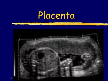
Placenta PowerPoint PPT Presentation
1 / 64
Title: Placenta
1
Placenta
2
Placenta Development
- Placenta
- Serves to protect the embryo as well as provide
nutrition, respiration, and excretion - Responsible for metabolism transfer of
nutrients, wastes, antibodies, hormones - Drugs infections can cross placenta
3
Placenta Development
- Develops from fetal chorion maternal
endometrium - Visualized at 8 to 10 weeks as a focal
homogeneous hyperechoic thickening - Fetal surface of placenta is smooth umbilical
cord is attached near center
4
Developing Placenta
5
Developing Placenta
6
Placenta Hormone Production
- Human Chorionic gonaditrophin (HCG)
- Prolactin
- Estrogen
- Progesterone
7
Evaluate the Placenta
- Reasons why we look at placenta
- R/O placenta previa
- R/O placenta abruption
- Localization prior to amniocentesis
- Placental aging
- Localization for CVS
8
Maternal Circulation
- The maternal (irregular lobular surface) surface
of the placenta (basilar plate - decidua basalis)
is divided into cotyledons, which can be thought
of as divisions of the placenta - Cotyledons - 10-38 functional lobes composed of
maternal sinusoids and chorionic villous
structures. - These cotyledons are formed by growth of septa
from the maternal endometrium into the placenta.
9
Maternal Circulation
- Each cotyledon contains maternal arterioles which
supply a pool of oxygenated blood into the
intervillous space which is found within the
placental lobules. - This intervillus space is formed by the villi
projections of the trophoblastic cells. These
trophoblastic cells serve as the fetal
capillaries.
10
Maternal Circulation
- Both maternal and fetal Blood flows over the
chorionic villi in the intervillous spaces. This
allows for exchange of oxygen nutrients. - This is the site of fetal-maternal exchange
- These fetal capillaries join into one fetal vein,
the umbilical vein, which feeds oxygenated blood
to the fetus. - After the blood circulates through the fetus the
deoxygenated blood exits the fetus via the two
umbilical arteries.
11
Maternal Circulation
- The two umbilical arteries break down into much
smaller arteries in the placenta where the blood
becomes oxygenated again. - Maternal blood reenters the maternal circulation
via basilar, subchorionic, interlobar, and
marginal veins (all which are maternal veins). - The rate of uteroplacental flow increase from 50
cc/min. at 10 wks to 500-600 cc/min. at term.
12
Maternal Circulation Review
- Maternal Circulation
- Placenta
- Cotyledons
- Maternal arterioles
- Pool of oxygenated blood
- Intervillous space
- Fetal capillaries
- Fetal vein
13
Placenta Vascularity
14
Placenta Vascularity
15
Fetal Circulation
- Oxygenated blood from the single umbilical vein
enters the fetal portal system through the
umbilical portion of the left portal vein. - From the left portal vein the blood has two
routes it can go through - Blood can go through the fetal liver and enter
the IVC through the hepatic veins - Or the blood can pass through the ductus venosus,
which bypasses the liver circulation and enters
the hepatic veins draining into the IVC.
16
Fetal Circulation
- The blood then goes from the left hepatic vein to
the IVC into the right atrium. - A small portion of blood from the right atrium
goes to the right ventricle to the lungs via the
pulmonary arteries. - But even most of this blood is shunted away from
the lungs and goes into the ductus arteriosus
that shunts blood directly into the descending
aorta for systemic circulation - After birth the ductus arteriosus constricts and
permanently closes becoming the ligamentum
arteriosum - , however, the majority of the blood is shunted
to the left atrium via the Foreman of ovali
17
Fetal Circulation
- The majority of blood in the right atrium goes to
the left atrium via the foreman ovali, which is a
hole between the right and left atrium. - The blood now goes from the left atrium to the
left ventricle to the aorta - Most of the blood then returns to the placenta
via two umbilical arteries this is deoxygenated
blood. - Some blood does remain to circulate within the
fetus
18
Review of Fetal Circulation
- 1. Umbilical vein (oxygenated blood)
- 2. Umbilical portion of hepatic portal
- 3. Left portal
- 4. Ductus venous
- 5. Left hepatic vein
- 6. IVC (Rt. Atrium, Lung's, Rt. Ventricle,
- 7. Foramen ovali
- 8. Lt. atrium 9. Lt. ventricle
- 10. Aorta
- 11. Two umbilical arteries
19
Fetal Circulation
20
Placenta
- A placenta is graded according to the following
- The amount and pattern of echogenic densities
within the placenta - The changes in the chorionic plate
- Chorion Plate This is the fetal surface- the
interface between the amniotic fluid the
placenta
21
Placenta (image)
22
Placenta
- Calcific deposits can occur in the substance of
the placenta throughout pregnancy - There is an increased incidence of placental
calcification with lower parity and higher
gestational age - Calcific deposits generally occur along the septa
and in the basalar plate
23
Grade 0 Placenta (Diaphragm)
24
Grade 0 Placenta
- Grade 0
- The chorionic plate is smooth, straight, and well
defined - The placenta has a homogeneous texture
- There are no focal densities seen in the basal
layer - Grade 0 placentas are usually seen in the first
and second trimester, prior to 28 weeks
25
Grade 0 Placenta
26
Grade 0 Placenta
27
Grade 0 Placenta
28
Grade 0 Placenta
29
Grade 1 (Diaphragm)
30
Grade I Placenta
- Characteristic undulations (indentation) of the
chorionic plate - Spotlike densities dispersed through out the
placental tissue (calcium deposits) - The basal layer is regular
- A Grade I placenta may appear as early as 14
weeks, but it is most commonly seen between 30-34
weeks and may persist until term
31
Placenta Grade 1
32
Placenta Grading (Grade l)
33
Grade I Placenta
- Few scattered calcifications
34
Grade II (Diaphragm)
35
Grade II Placenta
- Chorionic plate develops more marked indentations
- The placental substance is incompletely divided
by linear or comma shaped echogenic densities - Increase in the echogenic densities in the
placental substance - The basal layer also contains linear echogenic
densities - Indentations extending into the placenta but not
to the basal layer - Does not usually appear until after 30 weeks
36
Grade II Placenta (undulations)
37
Placental Clacifications (Grade II Placenta)
Calcifications are seen in aging placentas
38
Grade II Placenta
39
Grade III Placenta (Diaphragm)
40
Grade III Placenta
- The chorionic plate appears interrupted by
echogenic indentations that extend to basal layer - The placental substance is divided by these septa
and may contain anechoic or hypoechoic regions - Irregular echogenic areas throughout placenta
- Usually not seen until 35 weeks
- Found in 30 of term placentas
41
Grade III Placenta
- Echogenic foci along the basilar plate are more
prominent - Basal stripling - linear arrangement of echogenic
calcifications along the basilar plate, usually
not seen prior to 34 wks - Can be seen in the third trimester near term
- Prominent calcifications scattered throughout the
placenta and along the basalar plate
42
Grade III Placenta Cotelydons
ltgt
43
Grade III Placenta Cotelydons
44
Grade III Placenta Cotelydons
45
Placenta Grading Review (Grade 0-I)
- Grade 0
- No calcifications are seen, usually seen prior to
28 weeks - Grade 1
- Scattered calcifications are seen
- (Usually seen between 30-34 weeks)
- Can be seen prior to this and not be considered
abnormal
46
Placenta Grading Review (Grade II-III)
- Grade II
- Basal calcifications are seen
- 45 of placentas go to grade II
- More commonly seen over 30wks
- Grade III
- Basal and interlobar septal calcifications are
seen - Found in 30 of term placentas
- Not usually seen prior to 35 wks
47
Reasons the Placenta Ages to Quickly
- Hypertension - most common
- Smoking
- Sickle cell anemia
- Drug abuse
48
Reasons for Delayed Placenta Aging
- Rh sensitization
- Maternal diabetes
- Twins
49
Placenta Thickness
- 4-5 cm is a normal placental thickness.
- The placenta increases in thickness until 33
weeks of gestation, after that its thickness
gradually decreases - Anything greater than 6-7 cm or less than 4 cm is
abnormal - A mature placenta weighs 500 - 600 grams and is
16-20 cm in length
50
Reasons for a Thickened Placenta
- RH sensitization
- Maternal intrauterine infections
- Maternal diabetes
- Maternal anemia
- Feto-maternal hemorrhage
- Non-immune hydrops
- Chromosomal abnormalities
- Uterine contraction
51
Reasons for a Thin Placenta
- Preeclampsia
- Placental infarction
- Intrauterine infections
- Chromosomal abnormalities
- As a general rule of thumb, after 23 weeks the
placenta should be no thinner than 1.5 cm or no
thicker than 5.0 cm
52
Placenta
- Myometrium
- The muscle that forms the wall of the uterus.
- A contraction in the myometrium of the placenta
can cause it to appear thick or bulbous
53
Placenta
- Erythroblastosis fetalis
- A form of fetal anemia in which the fetal red
cells are destroyed by contact with a maternal
antibody that is produced by the mother in
response to a previous pregnancy - Severe fetal heart failure results
54
Placental Position
- Usually the placenta location is mid to fundal in
the uterus which reflects the site of
implantation. - Placenta positions
- 1. Posterior
- 2. Anterior
- 3. Lateral-right or left
- 4. Fundal
- 5. Combination
55
Placenta Positions
- Ultrasound Findings
- An over distended bladder focal uterine
contractions are the two most common technical
factors responsible for a false-positive
diagnosis by ultrasound
56
Placenta Migration
- The placenta is seen as a definite structure by
10-12 weeks and reaches its final location by 4
months - This term is used to describe the apparent shift
in the placental position early in pregnancy
prior to 16wks. - The placenta appears to be move from the cervical
to the fundal portion of the uterus.
57
Demonstrating the Lower Uterine Segment (LUS)
- 1. Demonstrate with a full bladder to R/O
- a. placenta previa
- b. complete previa
- If the placenta extends into the LUS, empty the
bladder because there may be a placenta previa. - Only with an empty bladder can you be sure the
cervix isn't falsely being lengthened by the
bladder being over distended. (Sagittal)
58
LUS
- Show that the placenta lies to one side of the
cervix. (Transverse) - It may be hard to demonstrate with a cephalic
presentation.
59
LUS
60
Translabial
61
Translabial ultrasound
62
TRANSLABIAL
63
TRANSLABIAL
64
Transvaginal

