
ABSTRACT - PowerPoint PPT Presentation
1 / 9
Title: ABSTRACT
1
ABSTRACT
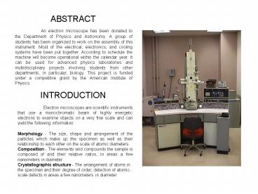
An electron microscope has been donated to the
Department of Physics and Astronomy. A group of
students has been organized to work on the
assembly of this instrument. Most of the
electrical, electronics, and cooling systems have
been put together. According to schedule the
machine will become operational within the
calendar year. It can be used for advanced
physics laboratories and multidisciplinary
projects involving students from other
departments, in particular, biology. This project
is funded under a competitive grant by the
American Institute of Physics.
- INTRODUCTION
Electron microscopes are scientific instruments
that use a monochromatic beam of highly energetic
electrons to examine objects on a very fine scale
and can yield the following information Morpholo
gy - The size, shape and arrangement of the
particles which make up the specimen as well as
their relationship to each other on the scale of
atomic diameters. Composition - The elements and
compounds the sample is composed of and their
relative ratios, in areas a few nanometers in
diameter Crystallographic structure - The
arrangement of atoms in the specimen and their
degree of order, detection of atomic-scale
defects in areas a few nanometers in diameter
2
BASIC COMPONENTS OF AN ELECTRON MICROSCOPE
Electron gun - The function of the gun is to
produce a fine beam of electrons of precisely
controlled energy (i.e. velocity) all coming
from a small source region
Lenses - A strong magnetic field is generated by
passing a current through a set of windings. This
acts as a convex lens, bringing off axis rays
back to focus. Focal length can be altered by
changing the strength of the current. C1 the
first condenser lens creates a demagnified image
of the gun crossover and C2 the second condenser
lens affects the convergence of the beam at the
specimen and the diameter of the illuminated area
of the specimen. The condenser aperture controls
the fraction of the beam which is allowed to hit
the specimen and therefore helps to control the
intensity of illumination, and in the SEM, the
depth of field. The objective lens forms an
inverted initial image, which is subsequently
magnified. The objective aperture is placed in
the back focal plane of the image and its
function is to select those electrons which will
contribute to the image, and thereby affect the
appearance of the image and to improve the
contrast of the final image. Magnification in
the electron microscope can be varied from
hundreds to several hundred thousands of times by
varying the strength of the projector and
intermediate lenses.
3
ELECTRICAL SYSTEM
The electrical system supplies power to all
components of the electron microscope. The
electrical system consists of four main
components. A single-phase 200/220/240Volt high
voltage power supply will be used for electron
beam acceleration in operation of the microscope.
A lens power supply will be used for the
electron lens excitation. A deflector coil power
supply will be used to deflect the electron beam.
Many sections of the microscope contain
miscellaneous circuits including a vacuum system
control circuit, a vacuum pump power supply, an
automatic exposure circuit, a camera drive
circuit, and an electron gun filament heating
circuit. These circuits are all solid-state
units to insure trouble-free operations and ease
of maintenance of the microscope.
4
(No Transcript)
5
CLOSED WATER SYSTEM
A closed water-cooling system is used in the
operation of this electron microscope. At a flow
rate of 4 Liters/min or more, the water system
will ensure that the electron microscope is kept
at safe operating temperatures. Pressurized
(1.5-5kg/cm2), cool, purified water is inserted
to the microscope while the warmer water is
expelled. The water cooler uses a refrigerant
compressor cycle to cool the incoming water.
Solenoid valves, manual valves, and constant
pressure valves control the pressure of the water
through the microscope depending on the pressure
and temperature of the water flow. Water is
carried from the water supply through the
water-cooling tank and into the microscope via ½
rubber tubing. The water enters under two paths.
In one path, water flows around the two oil
diffusion pumps, through the oil tank for the
objective lens, into the oil tank for the
condenser, intermediate and projector lenses, and
back out of the microscope kept at a constant
pressure of 3Liters/min. In the second path,
water flows through a thermoregulator, into the
column containing an objective lens, intermediate
lens, projector lens, condenser lens, and out of
the microscope at 1Liter/min. Filters are used
at junctions throughout the construction of the
closed water-cooling system to insure the purity
of the water flow.
6
THE VACUUM SYSTEM
Electrons are greatly influenced by the medium
through which they pass. Accordingly, it is
desirable that the pressure in the column be
maintained at 10-5 Torr or better. A poor vacuum
may cause high-tension electrical discharge,
specimen contamination, contrast reduction or
damage to the specimen itself. To achieve the
required vacuum degree, electron microscopes are
usually evacuated by oil rotary pumps and oil
diffusion pumps. The interior of the oil
rotary pump contains oil, which serves to
lubricate the pump and make it airtight. As the
rotor turns, the gas in one chamber is compressed
so that the pressure is greater than the
atmospheric pressure. This causes the gas to be
pushed out the exhaust valve into the atmosphere.
Simultaneously gas enters the other chamber for
the next compression stage. Since this is a two
stage pump, vibration and exhaust noise levels
are low. Unfortunately the rotary pump alone is
not enough to sustain a vacuum of 10-5 Torr, but
it is still useful for the initial pumping
procedure, and for maintaining the back pressure
of the oil diffusion pump.
7
THE VACUUM SYSTEM
- The oil diffusion pump is composed of a boiler
containing a heater, a water-cooled casing, and a
three-stage jet chimney. The oil heated by the
boiler enters the chimney as vapor, and is jetted
through a nozzle into the low pressure section at
supersonic speeds. This causes the gas molecules
from the column to diffuse into the oil jet
stream and the intermingled gas is compressed by
the kinetic energy of the jet flow and
transferred to the exhaust port. Finally the
jetted oil vapor is condensed by the water-cooled
casing and the condensed oil drains back into the
boiler. There is also a water-cooled baffle
which serves to prevent back streaming of oil
vapor. The oil diffusion pump cannot be operated
alone however, because the pressure on the
suction side must be approximately 1 Torr or
lesslower than the atmospheric pressure. Thus,
the oil rotary pump is used as an auxiliary pump
until the oil diffusion pump can be used. The
maximum obtainable pressure possible with this
system is approximately 10-8 Torr at a pumping
speed of several hundred liters per second
8
FUTURE PLANS
Work on the microscope will be performed chiefly
by the authors to ensure continuity of the
project into the next two academic years. These
students will be responsible for organizing
chapter participation next year, as well as
writing the final report in December. The
authors are registered for a Research
Participation class, which will guarantee at
least three hours per week per student involved.
Other interested members of the SPS Chapter will
be encouraged to participate in the assembly and
operation of the microscope, under supervision of
the authors or their successors. Incorporation
of the ASID system will eventually enhance the
visualization capabilities for imaging. It is
expected that after the assembly of the
microscope has been completed, it will be used to
promote multidisciplinary educational programs in
the Physics and Biology departments at Drake, in
order to ensure its continued use as an
educational tool. One possibility is the
development of research collaboration in
conjunction with the department of Microbiology,
in order to train undergraduate students in
techniques such as specimen preparation,
sectioning, staining, photography and analysis of
images.
9
(No Transcript)
Recommended
CrystalGraphics Presentations


