Eye Physiology - PowerPoint PPT Presentation
1 / 65
Title:
Eye Physiology
Description:
Because the different background intensities, the small squares do not ... For example, the American flag will not immediately appear red, white, and blue ... – PowerPoint PPT presentation
Number of Views:663
Avg rating:3.0/5.0
Title: Eye Physiology
1
Eye Physiology
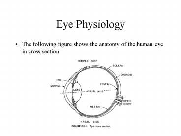
- The following figure shows the anatomy of the
human eye in cross section
2
Eye Physiology
- There are two types of receptors in the retina
- The rods are long slender receptors
- The cones are generally shorter and thicker in
structure - The rods and cones are not distributed evenly
around the retina. - Rods and cones operate differently
- Rods are more sensitive to light than cones.
- At low levels of illumination the rods provide a
visual response called scotopic vision - Cones respond to higher levels of illumination
their response is called photopic vision
3
(No Transcript)
4
(No Transcript)
5
Eye Physiology
- Rods are more sensitive to light than the cones.
6
Eye Physiology
- The eye contains about 6.5 million cones and 100
million rods distributed over the retina. - The density of the cones is greatest at the
fovea, this is the region of sharpest photopic
vision.
7
Eye Physiology
- There are three basic types of cones in the
retina - These cones have different absorption
characteristics as a function of wavelength with
peak absorptions in the red, green, and blue
regions of the optical spectrum.
- is blue, b is green, and g is red
- There is a relatively low sensitivity to blue
light - There is a lot of overlap
8
Eye Physiology
- The optic nerve bundle contains on the order of
800,000 nerve fibers. - There are over 100,000,000 receptors in the
retina. - Therefore, the rods and cones must be
interconnected to nerve fibers on a many-to-one
basis.
9
Contrast Sensitivity
0
1
2
3
4
Circle constant
Background constant
Just noticeable difference (JND) at 2
10
Contrast Sensitivity
0
1
2
3
4
Circle constant
Background constant
Just noticeable difference (JND) at 2
11
Contrast Sensitivity
0
1
2
3
4
Backgrounddifferent thenboth halves
Backgroundsame asright half
Just noticeable difference (JND) 4 (top) and
2 (bottom)
12
Contrast Sensitivity
0
1
2
3
4
Backgrounddifferent thenboth halves
Backgroundsame asright half
Just noticeable difference (JND) 4 (top) and
2 (bottom)
13
Contrast Sensitivity
- The response of the eye to changes in the
intensity of illumination is nonlinear - Consider a patch of light of intensity idI
surrounded by a background intensity I as shown
in the following figure
14
Contrast Sensitivity
- Over a wide range of intensities, it is found
that the ratio dI/I, called the Weber fraction,
is nearly constant at a value of about 0.02. - This does not hold at very low or very high
intensities - Furthermore, contrast sensitivity is dependent on
the intensity of the surround. Consider the
second panel of the previous figure.
15
Logarithmic Response of Cones and Rods
- The response of the cones and rods to light is
nonlinear. In fact many image processing systems
assume that the eye's response is logarithmic
instead of linear with respect to intensity. - To test the hypothesis that the response of the
cones and rods are logarithmic, we examine the
following two cases - If the intensity response of the receptors to
intensity is linear, then the derivative of the
response with respect to intensity should be a
constant. This is not the case as seen in the
next figure.
16
(No Transcript)
17
Logarithmic Response of Cones and Rods
- To show that the response to intensity is
logarithmic, we take the logarithm of the
intensity response and then take the derivative
with respect to intensity. This derivative is
nearly a constant proving that intensity response
of cones and rods can be modeled as a logarithmic
response. - Another way to see this is the following, note
that the differential of the logarithm of
intensity is d(log(I)) dI/I. Figure 2.3-1
shows the plot of dI/I for the intensity response
of the human visual system. - Since this plot is nearly constant in the middle
frequencies, we again conclude that the intensity
response of cones and rods can be modeled as a
logarithmic response.
18
Simultaneous Contrast
- The simultaneous contrast phenomenon is
illustrated below. - The small squares in each image are the same
intensity. - Because the different background intensities, the
small squares do not appear equally bright.
19
Simultaneous Contrast
- Perceiving the two squares on different
backgrounds as different, even though they are in
fact identical, is called the simultaneous
contrast effect. - Psychophysically, we say this effect is caused by
the difference in the backgrounds, but what is
the physiological mechanism behind this effect?
20
Simultaneous Contrast
- Perceiving the two squares on different
backgrounds as different, even though they are in
fact identical, is called the simultaneous
contrast effect. - Psychophysically, we say this effect is caused by
the difference in the backgrounds, but what is
the physiological mechanism behind this effect?
Lateral Inhibition
21
Lateral Inhibition
- Record signal from nerve fiber of receptor A.
- Illumination of receptor A alone causes a large
response. - Add illumination to three nearby receptors at B
causes the response at A to decrease. - Increasing the illumination of B further
decreases As response. - Thus, illumination of the neighboring receptors
inhibited the firing of receptor A. - This inhibition is called lateral inhibition
because it is transmitted laterally, across the
retina, in a structure called the lateral plexus.
22
Lateral Inhibition
23
Lateral Inhibition
- A neural signal is assumed to be generated by a
weighted contribution of many spatially adjacent
rods and cones. - Some receptors exert an inhibitory influence on
the neural response. - The weighting values are, in effect, the impulse
response of the human visual system beyond the
retina.
24
Lateral Inhibition
25
Mach Band Effect
- Another effect that can be explained by the
lateral inhibition. - The Mach band effect is illustrated in the figure
below. - The intensity is uniform over the width of each
bar. - However, the visual appearance is that each strip
is darker at its right side than its left.
26
Mach Band
- The Mach band effect is illustrated in the figure
below. - A bright bar appears at position B and a dark bar
appears at D.
27
Modulation Transfer Function (MTF) experiment
- An observer is shown two sine wave grating
transparencies, a reference grating of constant
contrast and spatial frequency, and a
variable-contrast test grating whose spatial
frequency is set at some value different from
that of the reference. - Contrast is defined as the ratio (max-min)/(max
min)where max and min are the maximum and
minimum of the grating intensity, respectively. - The contrast of the test grating is varied until
the brightness of the bright and dark regions of
the two transparencies appear identical.
28
Modulation Transfer Function (MTF) experiment
- In this manner it is possible to develop a plot
of the MTF of the human visual system. - Note that the response is nearly linear for an
exponential sine wave grating.
29
- Note that the envelope of the visible bars
generally follows the MTF curves of the previous
figure.
30
Logarithmic model for monochrome vision
- It has been postulated that the nonlinear
response of the eye to intensity variations is
logarithmic in nature and occurs near the
beginning of the visual information processing
system. - Below is a simple logarithmic eye model for
monochromatic vision.
31
Monochrome Vision Model
- The logarithmic/linear system eye model provides
a reasonable prediction of visual response over a
wide range of intensities. - However, at high spatial frequencies and at very
low or very high intensities, observed responses
depart from responses predicted by the model.
32
Lateral Inhibition
33
Extended model for Monochrome Vision
34
Subjective Color
- Intermittent pulses of white light are perceived
as colored light. - 1894 Benham invented the following experiment.
- Spinning the disk CCW
- Outer ring appears red
- Middle ring appears green
- Inner ring appears blue
- CW rotation reverses thecolors of the inner and
outer rings. - This effect is due to the temporal response of
the human visual system to flashing lights.
35
Chromatic Adaption
- The hue of a perceived color is dependent on the
adaption of a viewer. - For example, the American flag will not
immediately appear red, white, and blue if the
viewer has been subjected to high-intensity red
light before viewing the flag. - The colors of the flag will appear to shift in
hue toward the red complement, cyan.
36
(No Transcript)
37
Color Blindness
- Approximately 8 of males and 1 of females are
subject to some form of color blindness. - Monochromats only possess rods or rods plus one
type of cone. - Dichromats possess two of the three types of
cones. - Both monochromats and dichromats can distinguish
colors insofar as they have learned to associate
particular colors with particular objects.
38
Classroom Experiments
- Use Matlab to reproduce the Weber Fraction plot.
- Use Matlab to reproduce the Mach band effect with
uniform intensity bars (figure 2.3-2). - Use Matlab to reproduce the Mach band effect with
the sigmoid intensity profile (figure 2.3-2). - Use Matlab to reproduce the simultaneous contrast
phenomenon (figure 2.3-3). - Use Matlab to display a simple American flag and
a solid red square to reproduce the chromatic
adaption experiment from Pratt.
39
Color Vision Model
- Trichromatic Model of human color vision
- The eye possesses 3 types of sensors, each
sensitive over a different wavelength band - Receptor spectral sensitivities s1(l), s2(l), and
s3(l) represent the absorption pigments of the
retina. - Receptors produce signalswhere C(l) is the
spectral energy distribution of the incident
light source.
40
Color Vision Model
- Spectral Sensitivities si(l) in Fig 2.2-4 where
obtained by spectral absorption measurements of
cone pigments. - Direct physiological measurements are difficult
to perform accurately. - Indirect estimates of cone spectral sensitivities
have been obtained from measurements of the color
response of color blind individuals Konig and
Brodhun.
41
Color Vision Model
- The 3 signals e1, e2, e3 are subject to a
logarithmic transfer function and combined to
produce the outputs.
42
Color Vision Model
- Finally, the signals d1, d2, d3 pass through
linear systems with transfer functions H1, H2,
and H3, to produce the output signals g1, g2, g3
that provide the basis for perception of color by
the brain.
43
Color Vision Model
- d2 and d3 are related to the chromaticity of a
colored light. - d1 is proportional to luminance.
- This model satisfies the basic laws of
colorimetry. - If the spectral energy of a colored light changes
by a constant multiplicative factor, the hue and
saturation of light, as described by its
chromaticity coordinates, remains invariant over
a wide dynamic range, i.e., d2 and d3 do not
change. - The luminance d1 increases in a logarithmic
manner.
44
Color Vision Model
- Just like the monochrome model, the logarithmic
color vision model may be extend to a more
accurate model. - The linear transfer functions HE1, HE2, HE3,
account for the optical response of the eye - A point non-linearity is substituted for the
logarithmic transfer function.
45
Color Vision Model
- Sine wave response measurements for colored
lights were performed by van der Horst, de Weert,
and Bouman. - Chromatic response is shifted toward low spatial
frequencies relative to the luminance response.
46
Gamma Correction
- Red, green, and blue signals from video camera
sensors typically are linearly proportional to
the light striking each sensor. - However, light generated by CRT displays is
approximately equal to the display amplitude
drive signals raised to a power in the range of
2.0 to 3.0. - Gamma correction is a compensation process that
corrects for this nonlinear difference. - The camera sensor signal is passed through a
nonlinear system with a power, typically, of
about 0.45.
47
Gamma Correction
- In a video system, luminance of each of the
linear-light red, green, and blue (tristimulus)
components is transformed to a nonlinear video
signal by gamma correction, which is universally
done at the camera. - The Rec. 709 transfer function takes linear-light
tristimulus value (here L) to a nonlinear
component (here E'), for example, voltage in a
video system
48
Gamma Correction
- The linear segment near black minimizes the
effect of sensor noise in practical cameras and
scanners. Here is a graph of the Rec. 709
transfer function, for a signal range from zero
to unity
49
Gamma Correction
- An idealized monitor inverts the transform
- Real monitors are not as exact as this equation
suggests, and have no linear segment, but the
precise definition is necessary for accurate
intermediate processing in the linear-light
domain. - In a color system, an identical transfer function
is applied to each of the three tristimulus
(linear-light) RGB components.
50
Gamma Correction
- The nonlinearity of a CRT is a function of the
electrostatics of the cathode and the grid of an
electron gun it has nothing to do with the
phosphor. - The nonlinearity is a power function f (x)
xa, not an exponential function f (x) ex.
51
Gamma Correction
- To reduce TV receiver cost, gamma correction is
performed at the television camera rather than
the receiver. - A linear RGB image that has been gamma corrected
is called a gamma RGB image. - LCD displays are reasonably linear in the sense
that the light generated is approximately
proportional to the display amplitude drive
signal. - LCDs usually employ circuitry to compensate for
the gamma correction at the sensor.
52
Gamma Correction
- In video, a 0.45-power function is applied at the
camera - Synthetic computer graphics calculates the
interaction of light and objects. These
interactions are in the physical domain, and must
be calculated in linear-light values. It is
conventional in computer graphics to store
linear-light values in the frame buffer, and
introduce gamma correction at the lookup table at
the output of the frame buffer.
53
Gamma Correction
- If linear-light is represented in just eight
bits, near black the steps between codes will be
perceptible as banding in smoothly-shaded images.
This is the eight-bit bottleneck in the sketch. - Desktop computers are optimized neither for image
synthesis nor for video. They have programmable
"gamma" and either poor standards or no
standards. Consequently, image interchange among
desktop computers is fraught with difficulty.
54
References
- J.S. Lim, Two-Dimensional Signal and Image
Processing, Prentice Hall, 1991. - W.K. Pratt, Digital Image Processing, Wiley
Interscience, 3rd ed., 2001. - Charles Poynton's Frequently Asked Questions
about Colorhttp//www.inforamp.net/poynton/Color
FAQ.html - Charles Poynton's Frequently Asked Questions
about Gamma.http//www.inforamp.net/poynton/Gamm
aFAQ.html
55
(No Transcript)
56
Psychophysical Vision Properties
- Let E(l) represent the spectral energy
distribution of light emitted from some primary
light source. - Let t(l) and r(l) denote the wavelength dependent
transmissivity and reflectivity, respectively, of
an object - For a transmissive object, the observed light
spectral energy distribution is - C(l) t(l)E(l)
- and for a reflective object
- C(l) r(l)E(l).
57
Photometry
- The Commission Internationale de lEclairage
(C.I.E.) sets the standards for light and color. - SA is a tungsten filament lamp
- SB approximates direct sunlight
- SC approximates light from an overcast sky.
- A hypothetical source, called illuminant E, is
assumed to emit constant radiant energy at all
wavelengths.
58
Color Matching Quiz
- Is this Additive or Subtractive Color Matching?
59
Color Matching Quiz
- Is this Additive or Subtractive Color Matching?
60
Color Matching Quiz
- Is this Additive or Subtractive Color Matching?
61
Color Matching
- Additive Color Matching
Subtractive Color Matching
cyan
red
magenta
green
blue
yellow
62
Subtractive Color Matching
- The dye concentrations of the three spectral
filters are varied until a perceptual match is
obtained with a reference white W. - The dye concentrations are recorded as A1 (W), A2
(W), A3(W).
63
Subtractive Color Matching
- Next, the dye concentrations of the three
spectral filters are varied until a perceptual
match is obtained for the colored light C. - If a match is possible, record the intensities
asA1 (C), A2 (C), A3(C). - The tristimulus values are computed as
64
Subtractive Color Matching
- One primary P3 is superimposed with the light
C and compared to the overlap of the other two
primaries P1 and P2. - All primaries are adjusted until there is a
match. - If a match is possible, the tristimulus values
are computed as
65
Subtractive Color Matching
- Two primaries P2 and P3 are superimposed with
the light C and compared to the other primary
P1. - All primaries are adjusted until there is a
match. - If a match is possible, the tristimulus values
are computed as


