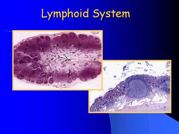
Lymphoid System PowerPoint PPT Presentation
1 / 32
Title: Lymphoid System
1
Lymphoid System
2
Lymphoid System
- Functions
- Immunologically mediated defense against foreign
antigens (often infectious) - Intimately associated with vascular system
3
Components of lymphoid system
- Lymph and lymph nodes
- Lymph fluid scavenged from the intersitium
- Collected in thin walled lymphatic vessels
- Returned to general circulation
- Contains leukocytes
- Filtered through small specialized organs (lymph
nodes)
4
Additional lymphoid Tissues
- Thymus
- Spleen
- Tonsils
- Mucosal associated lymphoid tissue (MALT)
- Gut-associated (GALT)
- Bronchial-associated (BALT)
- Bursa of Fabricius (avian)
5
Lymph node
- Locations
- Distributed along regional lymphatics
- Aggregates in
- Neck, axillae, groin, lung hilus
- At sites were major lymphatic vessels converge
6
Lymphatic vessels
- Thin walled
- Originate as blind open vessels
- Have valves to allow one-way flow
- Muscular activity is necessary for efficient flow
7
8
Lymph node
- Functions
- Filtration of lymph
- Initiation of immune response
- Activation and proliferation of lymphocytes
9
Lymph node
- Cell types
- Lymphocytes and plasma cells
- Accessory cells
- Macrophages
- Follicular dendritic cells
- Interdigitating dendritic cells
- Stromal cells
10
Lymph node
- Circulation of lymphocytes and lymph
- Enter and exit via blood or lymph
- If able to respond to any antigen presented
stay and differentiate - If unable to respond exit via efferent
lymphatics
11
Lymph node architecture
12
Lymph node architecture
HEV specialized Post-capillary
venules. Vascular addressins Special surface
markers Cuboidal endothelium
13
Compartments
- Sinuses
- Lined by endothelium and macrophages
- Contain lymphocytes, macrophages, langerhans
cells - Site of filtration and antigen processing
14
Lymph node capsule
15
Lymph node - subcapsular sinus
16
Compartments
- Vascular system
- Entry/exit site for lymphocytes and dendritic
cells
17
Postcapillary venules
18
Compartments
- Interstitial tissue
- Cortex
- Paracortex
- Medullary cords
- Site of maturation and proliferation of
lymphocytes
19
Lymph node
20
Lymph node reticulum stain
21
Lymph node
22
Lymphoid follicle
23
Lymphoid follicle immunohistochemistry
T-cell marker
B-cell marker
24
Follicular dendritic cells
25
Antigen processing cell nucleus
26
Lymph node medullary cords
27
Medullary cords reticulum stain
28
Lymph node medullary sinuses
29
Plasma cell
30
Pig lymph node
- Inverted structure
- Cortex is internal
- Medullary tissue is external
- Still has diffuse paracortical tissue and nodular
(follicular) tissue
31
Compartmentalized Responses
- Cell-mediated responses will expand the
paracortex - Humoral responses will expand the cortex
32
Hemal nodes
- Associated with blood vessels not lymphatics
- Are blood filled
- Have capillary plexuses with post-capillary
venules similar to HEV - Have cortex and medulla (nodular and diffuse
tissue - Are little spleens

