Cell Division PowerPoint PPT Presentation
1 / 35
Title: Cell Division
1
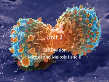
Unit 2
- Cell Division
- Mitosis and Meiosis I and II
2
Mitosis Word Meanings
- Chromosome
- Etymology Greek Chromo colored soma body
- Centromere
- Centro center mere part
- Inter
- Etymology Latin between
- Pro-
- Etymology Greek before
- Meta-
- Etymology Greek after
- Ana-
- Etymology Greek up
- Telo-
- Etymology Greek end
- Cytokenesis
- Etymology Greek Cyto cytoplasm kenesis
movement
3
- Reasons why a cell needs to divide.
- Destroyed cells need to be replaced.
- Cell size
- Most living cells are between 2 and 200
micrometers in diameter (3 feet 1 meter 1
million micrometer) - Limitation of cell size
- Surface area to volume ratio
- Increase cell size will increase volume of cell
- As cells grow, the amount of surface area becomes
too small to allow materials to enter leave the
cell quickly enough by diffusion. - Diffusion (see unit 1)
- fast efficient over short distances
- If cell gets too large, would decrease diffusion.
4
- The nucleus
- DNA in the nucleus is copied to make proteins
which run the cells activities in the cytoplasm. - Cell size is limited by the amount of proteins
the DNA is able to make to control the cell.
5
- DNA Packing (occurs before cells divide)
- DNA is a very long thin molecule (cant see it)
- In order to fit in the nucleus it has to get
really compact ( packed in closely). - The DNA does this by wrapping around proteins
called Histones, and folding in on itself - This is known as Chromatin. (We can see this)
- When Chromatin is wrapped up on itself and takes
on a distict shape (looks like an X) it is
known as a Chromosome (We can see this too)
6
- Chromosomes
- Human cells have 46 Chromosomes
- Humans get 23 from the mother and 23 from the
father. - Before the DNA wraps up into a chromosome, the
DNA gets copied. - Before cell divides each DNA piece winds up into
a csome - One side of the chromosome is the original
chromosome - The other side of the chromosome is the copy of
the original chromosome. - When a Chromosome is paired we call each side a
Chromatid. (You have 2 Chromatids for each
Chromosome) - Chromosomes pairs are held together by a
centromere. - When the chromatids separate into separate nuclei
we call them chromosomes again.
Chromatids
Centromere
7
After DNA Replication2 pairs per type of
chromosome
- Before DNA Replication
- 1 pair per type of
- chromosome
Homologous Pairs
- Individual chromosomes
- 1 from each parent
Dad
Mom
8
- Cell Cycle
- Interphase before cell division
- G1 phase
- cell growth
- production of new organelles
- G0 (G zero)
- period when cell is not preparing to divide.
- Here cells lack growth factors
- Cells stay in the G0 until there is a reason for
them to divide. - Ex. Muscle, brain, nerve cells
- S phase Double the DNA
- DNA replication (duplicate)
- Go from 46 to 92 single chromosome molecules per
cell in humans - G2 phase
- cell prepares to divide
9
- M phase Mitosis Cell Division
- Prophase
- chromosomes shorten, thicken, and become visible
- nucleus and other organelles break down and
disappear - Organelles called centrioles begin to make
spindle fibers
10
- Metaphase
- spindle fibers become present and attach to
centromeres of chromosome pair. - chromosomes line at center of cell (metaphase
plate equator)
11
- Anaphase
- The Chromatids are pulled apart towards polar
ends of the cell.
12
- Telophase
- New nuclei form around each set of chromosomes
- two new cells start to take shape
- chromosomes begin to make proteins
13
- Cytokinesis
- In animal cells
- Animal cells undergo cytokinesis by pinching off
along their equator - This works because their membrane is flexible.
14
- In plant cells
- Plant cells cannot do the same as animal cells
because of their cell wall. - Plant cells send vesicles filled with cell wall
material to their equator where they fuse
together. - This fusion creates cell plates that fuse with
the cell wall separating the two cells.
15
- Cancer
- It is the disease where the bodys cells DO NOT
STOP mitosis. - Results in tumors forming throughout the body.
- A tumor is a bunch of cells in one area of the
body that keep dividing. - These cells can then move on to other parts of
the body causing tumors to develop in other
regions. - Cancer kills by causing your organs to
malfunction.
16
- Cell Differentiation
- Unspecialized cells develop into mature forms and
functions - Each cell has a full set of DNA
- BUT, each type of cell uses only specific genes
on the DNA to carry out its functions - Stem Cells
- Can develop into a variety of cell types
- Will divide by mitosis into either two stem
cells, or, a stem cell and a specialized cell.
17
(No Transcript)
18
Mitosis Pop Quiz
1.___________________________
2._________________________
4.__________________________
3.___________________________
19
Meiosis Word Meanings
- Homologous
- Etymology Greek homologos to agree
- having the same position, value, or structure
- Tetrad
- Etymology Greek
- a group or arrangement of four
- Dyad
- Etymology Late Latin dyas, from Greek, from dyo
- pair two individuals
- -ploid
- having or being a chromosome
- Di-
- Etymology Latin, from Greek akin to Old English
twi- - twice two double
- Ha-
- Etymology Greek single
20
- Somatic cells and Gametes
- Somatic cells are body cells.
- Make up most of your body cells.
- These are diploid (2n)
- Have two sets of chromosomes.
- 1 set from father 1 set from mother
- Gametes sex cells(sperm/egg cells)
- These are haploid (n)
- Have only one set of chromosomes
- Types of Chromosomes
- Autosomes contain genes not associated with sex
- Sex chromosomes directly control sexual traits.
21
Meiosis The creation of sex cells
- Interphase (G1, S, G2)
- All your cells start off as diploid cells
- Diploid (2n) cell with two of each kind of
chromosome (In humans 46 23 from each parent) - G1 Cell grows DNA is long and stringy and is
not visible. (so the DNA can be copied) - S DNA gets replicated (copied).
- G2 Cell continues growing and prepares to
divide.
22
Meiosis I
- Prophase I
- The chromatin wind-up, creating chromosomes which
can now be seen. - Centriols create Spindle Fibers
- Nuclear envelope disappears
- Each chromosome pair then actively seeks out its
homologous chromosome pair. - Homologous pairs are about the same size and
shape - This is called a tetrad (four chromatids)
- Crossing Over will now happen.
- where two non-sister chromatids exchange genetic
material.
23
After DNA Replication2 pairs per type of
chromosome
- Before DNA Replication
- 1 pair per type of
- chromosome
Homologous Pairs each individual chromosome has
been copied
- Individual chromosomes
- 1 from each parent
Dad
Mom
24
Prophase I
25
- Metaphase I.
- The spindle fibers attach to the centromere on
each chromosome pair. - Homologous chromosomes are lined up on the
metaphase plate (equator) by the spindle fibers. - 2 rows of 23 chromosome pairs 46 pairs total
26
- Anaphase I
- The tetrad gets pulled apart homologous
chromosomes separate. - CHROMATIDS DO NOT GET PULLED APART YET!
- Each homologous chromosome moves to opposite
poles.
27
- Telophase I / Cytokenesis
- Individual nuclear envelopes begin to surround
the separate chromosome pairs - Cytokenesis separates the one cell in two.
28
- END OF MEIOSIS I
- Two cells are created each with 23 chromosome
pairs
29
Meiosis II
- Prophase II
- Each dyad (2 Chromatids 1 Chromosome pair) are
connected by a centromere. - Nuclear envelope disappears
- The centrioles create spindle fibers again.
30
- Metaphase II
- The spindle fibers attach to the centromere on
each dyad (1 Chromosome pair 2 Chromatids). - The dyads are lined up on the metaphase plate
(equator) by the spindle fibers. - 1 row of 23 chromosome pairs in each cell.
31
- Anaphase II
- The individual sister chromatids from each dyad
get pulled apart by the spindle fibers - Each sister chromatid ends up on opposite poles
of the cell
32
- Telophase II / Cytokenesis
- The shape of the cell changes, beginning to form
two cells. - New nuclei form around each set of chromosomes
- The cytoplasm of both cells divides once again.
- Four gametes ( sex cells) are now created
33
- END OF MEIOSIS II
- Four unique haploid cells are created, each with
a half set of chromosomes compared to the
original (Original Parent cell had 46, each
daughter cell has only 23 chromosomes). - Haploid (n) cell with only 1 of each kind of
chromosome (in humans 23 chromosomes)
34
- Mitosis Chromosome Number Flow
Interphase
Mitosis
46 chromosome pairs
46 individual chromosomes 2n
46 individual chromosomes 2n
46 individual chromosomes 2n
35
- Meiosis I II Chromosome Number Flow
Meiosis I
Interphase
46 individual chromosomes 2n
46 chromosome pairs
23 chromosome pairs
23 chromosome pairs
Meiosis II
23 individual chromosomes n
23 individual chromosomes n
23 individual chromosomes n
23 individual chromosomes n

