PowerPoint Presentation Presentazione di PowerPoint - PowerPoint PPT Presentation
1 / 1
Title:
PowerPoint Presentation Presentazione di PowerPoint
Description:
... After hybridization, ligation and amplification according to the instructions of ... Recently a Multiple Ligation of Probe Amplificatio (MLPA) kit, covering 33 of ... – PowerPoint PPT presentation
Number of Views:111
Avg rating:3.0/5.0
Title: PowerPoint Presentation Presentazione di PowerPoint
1
NOVEL GENOMIC REARRANGEMENTS OF THE ATM GENE
IDENTIFIED IN ATAXIA-TELANGIECTASIA
ITALIAN PATIENTS
Simona Cavalieri 1, Ada Funaro 1, Paola Porcedda
2, Valentina Turinetto 2, Nicola Migone 1,
Patrizia Pappi 1, Richard Gatti 3, Alfredo Brusco
1
1 Department of Genetics Biology and
Biochemistry, University of Torino, and Medical
Genetics Unit, S. Giovanni Battista Hospital,
Torino, Italy 2 Department of Clinical and
Biological Sciences, University of Turin, Regione
Gonzole 10, Orbassano, Italy 3Department of
Pathology and Laboratory Medicine ,The David
Geffen School of Medicine at UCLA, Los Angeles,
California, USA.
Correspondence to alfredo.brusco_at_unito.it
Mutations identified in 19 Italian A-T Patients
INTRODUCTION Mutations in the ATM gene lead to
loss-of-function alleles. The most common
mutations are nonsense, splice-site variants,
small insertions or deletions and missense. Large
genomic deletions (LGDs) are rare but can lead to
the same phenotype. Here we report a multi-step
mutation screening strategy on the ATM gene in 19
unrelated Italian A-T patients which allowed us
to identify 37 of the 38 expected defective
alleles.
RESULTS (1) Multi-step mutation analysis
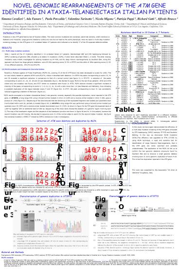
Table 1 reports all the 37 mutations identified
in 19 unrelated Italian A-T patients.
Standardized SNP and STR haplotyping followed by
DHPLC screening of genomic DNA, allowed us to
detect 31 mutations (?82), 9 were never
described (Table 1, in bold). The other missing
mutations were initially investigated for
splicing mutations by RT PCR, and for deep
intronic rearrangements by Southern Blot. Using
this approach we found two large genomic
deletions one of 8.5kb spanning exons 32-36 in
AT9TO and the other of 18kb spanning exons 21-29
in two different patients, AT21TO and AT23TO.
- MLPA analysis and breakpoint characterization
- Recently a Multiple Ligation of Probe
Amplificatio (MLPA) kit, covering 33 of the 65
ATM exons has been developed to screen for LGDs.
This kit was initially tested on patients AT9TO
and AT21TO, where it identified both deletions.
In AT9TO the peaks corresponding to exons 33, 35
and 36 revealed a significant reduction in
comparison to that of a normal control (see
figure 1) in AT21TO a reduction of the peaks
corresponding to exons 21, 24, 25, 28 and 29 was
detectable (fig.1c). We decided to apply MLPA to
the last three patients AT34 and AT39 where only
one mutation was found, and AT37 where no
mutations were found. In AT39, MLPA analysis
showed a significant increase of intensity of the
peaks corresponding to exons 4, 7, 8, 10, 12, 16,
19 with a ratio more than 1.4 (see Materials and
Methods), thus indicating a probable duplication
of the region between exons 4 and 19 (figure 2b).
In AT37, the peak corresponding to exon 31 was
consistently reduced suggesting a deletion of
that exon (figure 2c). - MLPA results prompted us to better
characterize these 2 new genomic variants,
especially the possible duplication, never
reported in the ATM gene. In AT39TO, a long-range
PCR using a forward primer located in intron 19
(19F) and a reverse primer located in intron 5
(4R), gave a product of 1.9kb (figure 3c).
Sequencing of this band showed that the beninning
of the duplicated fragment was located just
upostream exon 4 and ended within exon 20,
partially truncated (figure 3d). In AT37TO a
long-range PCR was performed using a forward
primer located just upstream exon 30 (30F) and a
reverse primer located downstream exon 31 (31R).
As shown in figure 4a the PCR gave the expected
band of 1,987 bp together with an additional band
of 956 bp. Sequencing of the lower band showed
the deletion of a genomic region involving exon
31.The map of the breakpoint region and the
sequence of the breakpoint junction are shown in
figure 4b and 4c. In this patient, however the
second mutation was still missing. We decided to
sequence exon 31 of the normal allele to search
for the second hidden mutation. We found the
nonsense mutation c.4396CgtT missed by DHPLC
analysis as it was in emozigosys.
Table 1 Letters were assigned to each haplotype
associated to a different mutation. Recurring
haplotypes are grey shaded.Mutations
corresponding to affected haplotypes are reported
. New mutations are in bold. Superscripts a
first allele b second allele h homozygote
patient. FSframeshift T truncation
Ababerrant splicing.
Detection of ATM exon deletions and duplication
by MLPA
CONCLUSIONS In this study we have again
demonstrated the efficiency of a multi-step
mutation screening of the ATM gene composed by
STR haplotyping, DHPLC analysis, RT-PCR and
Southern Blotting. In this way we discovered
35/38 mutations (92).This efficiency was
upgraded to 97 (37/38) by using MLPA technique,
a rapid and sensitive test for identification of
Large Genomic Rearrangements, that in the ATM
gene are rarely searched and probably
understimated. The application of the MLPA kit
led us to confirm the two genomic deletions
previously found by Southern Blot and to discover
a small genomic deletion involving exon 31 and a
genomic duplication of exons 4-20. This is the
first duplication reported in the ATM
gene. This work was supported by the
Association Gli Amici di Valentina Grugliasco,
Italy.
A
18
33
35
36
21
24
25
31
CTRL
CTRL
12
28
7
19
16
6
29
4
8
10
AT39TO
AT39TO
33
AT9TO
18
6
7
8
12
35
36
19
16
4
10
B
AT21TO
AT37TO
31
C
21
24
25
28
29
Figure 1
Figure 2
Figure 1A and 2A MLPA chromatograms
representing a normal control. Each peak
represents an exon recognized by the ATM-specific
probes. In AT9, AT21 and AT37 the peaks
representing the deleted exons are indicated by
red arrows. In AT39 arrows indicate the
duplicated exons. Note Exon deletions are
apparent by a 40 reduction in peak area of a
specific probe exon duplications are showed by a
40 increment in peak area. For AT39, the maximum
value of peak height has been increazed respect
to the others in order to better appreciate the
difference.
Characterization of genomic deletion in AT37TO
Figure 4
Characterization of genomic duplication in AT39TO
Figure 3
Material and Methods Total genomic DNA
extraction, STR haplotyping, DHPLC analysis,
RT-PCR and Southern Blot analysis have been
detailed described in Cavalieri et al. Human
Mutation (mutation in brief) 925, 2006. MLPA
analysis A total of 100ng of genomic DNA was
used as a starting material with the SALSA MLPA
KIT P123 ATM available from MRC Holland
(www.mrc-holland.com). The probe mix included in
this MLPA kit contains probes of 33 of the 65
exons, as well as control probes for sequences
located in other genes.After hybridization,
ligation and amplification according to the
instructions of the manifacturers, 1ulof the PCR
products were mixed with 0.2ul of ROX-labeled
internal size standard, separated on ABI Prism
3100Avant automatic sequencer (applera) and
analyzed using the Gene Scan software. For data
analysis, the peaks sizes and areas were
transferred to an Excel file. For normalization,
relative probe signals were calculated by
dividing each measured peak area by the sum of
all peak areas of that sample. The ratio of each
relative probe signal from patients compared to a
control sample was then calculated. An exon
deletion was considered when the ratio was lower
than 0.7 while a duplication when the ratio was
more then 1.3.


