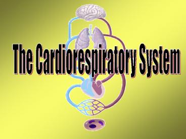
The Cardiorespiratory System PowerPoint PPT Presentation
1 / 16
Title: The Cardiorespiratory System
1
The Cardiorespiratory System
2
The Vascu
lar System
Veins and Venules
- Carries blood towards the heart
- Limited contractibility elasticity
- One-way valves
Arteries and Arterioles
- Carries blood away from the heart
- thick smooth muscle wall
- Contractible elastic
The Cardiorespiratory System
3
General Characteristics of the Heart
- Location Mid left chest
- Functions as a double pump
Right side deoxygenated blood
Left side oxygenated blood
The Cardiorespiratory System
4
The Heart's Linings
- protective sac
surrounding heart, and filled with fluid to
reduce friction
Pericardium -
Epicardium -
- outer lining of the
heart
Myocardium -
the heart muscle
Endocardium -
the inner lining of the
heart
The Cardiorespiratory System
5
Structure of the Heart (Anterior)
Aorta
Left Pulmonary artery
Superior Vena Cava
Branches of Left Pulmonary artery
Branches of Right Pulmonary artery
Pulmonary trunk
Left Pulmonary veins
Right Pulmonary veins
Left Atrium
Right Atrium
Branch of Left Coronary Artery
Right Coronary Artery
Great Cardiac Vein
Small Cardiac Vein
Right Ventricle
Left Ventricle
Thoracic aorta (descending)
Inferior Vena Cava
The Cardiorespiratory System
6
Chambers of the Heart
Two Atria
- Right atrium gets deoxygenated blood from the
superior and inferior vena cava
- Left atrium gets oxygenated blood from pulmonary
veins
Two Ventricles
- Right pumps blood to lungs to get oxygenated
- Left has thicker wall and pumps to the body
Ventricles are separated by interventricular
septum
The Cardiorespiratory System
7
The Four Valves of the Heart
Atrioventricular valves (gateway to ventricles)
- Right tricuspid
- Left bicuspid
- Cusps attached to papillary muscles by chordae
tendinae
- Leaks murmurs
Semilunar valves (gateway to lungs and aorta)
- Pulmonary and aortic
The Cardiorespiratory System
8
Internal Struc
tures of the Heart
The Cardiorespiratory System
9
Internal Struc
tures of the Heart
Right Side
Left Side
- Superior and inferior vena cava
- Right atrium
- Right ventricle
- Pulmonary artery
- Tricuspid valve
- Pulmonary valve
- Aorta and thoracic (descending) aorta
- Left atrium
- Left ventricle
- Pulmonary vein
- Bicuspid (mitral) valve
- Aortic valve
Common structures of both Chordae tendinae,
Papillary muscles, and the Interventricular septum
The Cardiorespiratory System
10
Path of Blood
through the Heart
Right Side
Left Side
- 7. Pulmonary veins
- 8. Left atrium
- 9. Mitral valve (Bicuspid)
- 10. Left ventricle
- 11. Aortic valve
- 12. Aorta
- Gas exchange with working cells
- Superior and inferior vena cava
- 2. Right atrium
- 3. Tricuspid valve
- 4. Right ventricle
- 5. Pulmonary valve
- 6. Pulmonary artery
- Gas exchange in lungs
12.
8.
5.
11.
9.
10.
4.
3.
11
Pulmonary and Systemic Blood Flow
1. Deoxygenated blood (venous blood) flows from
the upper body through the superior vena cava and
lower body through the inferior vena cava.
2. Right atrium receives venous blood.
3. Venous blood moves through the tricuspid
valve.
4. Venous blood is in the right ventricle.
5. Venous blood is pumped through the
semi-lunar valve of the pulmonary trunk
Tricuspid valve closes.
6. Venous blood travels from the right and left
pulmonary arteries to the lungs.
Gas exchange in the lungs
The Cardiorespiratory System
12
Pulmonary and Systemic Blood Flow
7. Oxygenated blood travels from the lungs
through the left and right pulmonary veins.
8. Oxygenated blood empties into the left
atrium.
9. Oxygenated blood passes through the bicuspid
valve.
10. Oxygenated blood is in the left ventricle.
- Left ventricle pumps oxygenated blood through
semi- - lunar valve of aorta Bicuspid valve closes.
12. Oxygenated blood is in the aorta.
13. Oxygenated blood is sent into the systemic
circulation through the brachiocephalic, left
common carotid, subclavian arteries and thoracic
aorta to the rest of the body.
The Cardiorespiratory System
Gas exchange with working cells
13
Myocardium
The myocardium or heart muscle is responsible for
the contractions that force blood out of the
heart and around the body.
Characteristics of myocardium include
- involuntary contraction
- striated (actin and myosin)
- contractions are activated by calcium and follow
the sliding filament theory of contraction - cells are connected via intercalated discs
(leaky membranes) that allow electrical signals
to pass between cells resulting in functional
synctium (all cells contracting in unison) - atrial cells are separated from ventricle cells
- cells (fibres) are similar to slow twitch muscle
fibres (large of mitochondria capillaries)
highly aerobic in nature
The Cardiorespiratory System
14
Conduction System of the Heart - SA node
Sinoatrial node (SA) specialized area of tissue
located in the posterior, superior right atrium
of the heart responsible for initiation of
electrical signals proceding heart contraction
(pacemaker)
SA node acts both as muscle and nerve tissue
- contracts (muscle)
SA node
- transmits impulse (nerve)
- the SA node sets the rate of contraction of the
heart
- the autonomic nervous system regulates it
The Cardiorespiratory System
15
Conduction System of the Heart
- SA node sends electrical signal through right and
left atria
2. Both atria contract simultaneously (top down)
forcing blood to the ventricles
- Signal then moves to inferior atria to the AV
(atrioventricular node)
4. After delay of 0.1sec, AV node transmits
signal through the Bundle of His
(interventricular septum)
5. Signal travels through to Purkinje fibres in
the myocardium of the ventricles.
6. Ventricles contract from the bottom up to
push blood into the pulmonary arteries and the
aorta.
The Cardiorespiratory System
16
Electrocardiogram
QRS complex electrical signal for contraction
throughout the ventricles, ventrical contraction
and atrial repolarization.
P wave spreading of electrical signal for
contraction throughout the atria and atrial
contraction.
T wave ventricle resets itself for the next
contraction (repolarization).
The Cardiorespiratory System

