Transport/Circulatory System - PowerPoint PPT Presentation
Title:
Transport/Circulatory System
Description:
... Provides oxygenated blood to rest of body Evolution of circulatory system fish amphibian reptiles birds & mammals A ... Why is it an advantage to get big ... – PowerPoint PPT presentation
Number of Views:144
Avg rating:3.0/5.0
Title: Transport/Circulatory System
1
Transport/Circulatory System
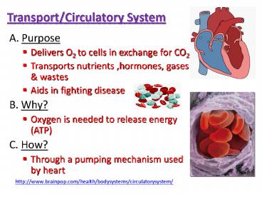
- A. Purpose
- Delivers O2 to cells in exchange for CO2
- Transports nutrients ,hormones, gases wastes
- Aids in fighting disease
- B. Why?
- Oxygen is needed to release energy (ATP)
- C. How?
- Through a pumping mechanism used by heart
http//www.brainpop.com/health/bodysystems/circula
torysystem/
2
D. Pathway of Blood -2 part closed circulatory
system
- 1.Pulmonary Circulation
- (Right Side of )
- Takes deoxygenated blood to the lungs returns
oxygenated blood back to heart - 2. Systemic Circulation
- (Left side of )
- Provides oxygenated blood to rest of body
3
Evolution of circulatory system
Not everyone has a 4-chambered heart
fish
amphibian
reptiles
birds mammals
2 chamber
3 chamber
3 chamber
4 chamber
V
A
A
A
A
A
A
A
V
V
V
V
V
4
E. Pathway
S. VENA CAVA
O2 IN
RIGHT ATRIA
RIGHT VENTRICLE
PULM ARTERY
BOTH LUNGS
PULM. VEIN
I. VENA CAVA
LEFT ATRIA
CO2 OUT
LEFT VENTRICLE
AORTA
BODY
BACK TO VENA CAVA
5
F. Structure of the Heart 4 chambered
- Right side carries O2 poor (deoxygenated) blood
to lungs - Left side carries O2 rich (oxygenated) blood to
the rest of the body - Analogous to cytoplasm of one celled organisms
A aorta largest arteryB pulmonary arteries (deoxygenated blood)C pulmonary veins (oxygenated blood)D left atrium upper chamber (thin)E valve prevent backflowF left ventricle lower chamber (thick)G right ventricle H valve I vena cavae J right atrium (areas shaded red have oxygenated blood while those shaded blue have deoxygenated blood)
Left side
Right side
6
Diagram of Heart and Blood Flow
- http//www.youtube.com/watch?vKSbbDnbSEyM
- http//www.ask.com/youtube?qcirculatorysongvide
oclipvq0s-1MC1hcE
7
Figure 37-5 The Three Types of Blood Vessels
BLOOD VESSELS http//www.youtube.com/watch?vCjN
KbL_-cwA
Section 37-1
Vein
Artery
Capillary
8
G. Blood Vessels
- Arteries Carry oxygenated blood away from the
heart high pressure - 2) Veins Carry deoxygenated blood to the heart
have valves to prevent backflow low pressure - Capillaries tiny, tiny vessels where gas
exchange occurs connect arteries to veins
9
H. Blood Composition - human body contains 4-6
liters
Part(s) Description Diagram Disease
Plasma (liquid) transporting nutrients and hormones 90 water, 10 other Yellow color
RBC (erythrocytes) Made in bone marrow disk-shaped lack nuclei Carries O2 using hemoglobin (allows RBCs to carry O2 ). Anemia (lack of iron) Sickle cell disease
Platelets cell fragments, not cells Blood clotting Dot-like fragments scattered Hemophilia (blood doesnt clot)
WBC (leukocytes) Functions in the immune system by attacking foreign substances Larger than RBCs Has a nucleus Leukemia (too many abnormal WBCs are produced)
10
Leukemia Smear
SICKLE CELL ANEMIA
Diseased white blood cell
11
Types of WBCs
Copy these notes under chart
- 1.Phagocytes
- Engulfs and destroy bacteria
- 2.Lymphocytes
- Produce antibodies that clump antigens (bacteria)
12
Figure 37-7 Blood
- Centrifugation separating the parts of blood
into layers based on density
Section 37-2
Plasma
Platelets
White blood cells
Red blood cells
Whole Blood Sample
Sample Placed in Centrifuge
Blood Sample That Has Been Centrifuged
13
Figure 37-7 Blood
Section 37-2
Plasma
Platelets
White blood cells
Red blood cells
Whole Blood Sample
Sample Placed in Centrifuge
Blood Sample That Has Been Centrifuged
14
Figure 37-7 Blood
Section 37-2
Plasma
Platelets
White blood cells
Red blood cells
Whole Blood Sample
Sample Placed in Centrifuge
Blood Sample That Has Been Centrifuged
15
Clotting Process - uses platelets How Does blood
clot http//www.bing.com/videos/search?qclotting
processanimationqsASskAS1FORMQBVRpqclotti
ng20processsc8-16sp2qsASskAS1adltstrict
viewdetailmidBB4338AF0FA5F3276DCDBB4338AF0FA5F
3276DCD
2. Platelets clump sealing the hole
1. Break in capillary wall
3. Protein fibersbuild the clot
16
Heart Disease (Basics 1)
- Video Understanding Heart Disease (Basics 1)
http//www.bing.com/videos/search?qHEARTDISORDER
SVIDEOFORMVIRE14adltstrictviewdetailmid8B
44B4B68E548634E8A58B44B4B68E548634E8A5
Heart disease death rates 1996-2002Adults ages
35 and older
17
Women Heart Disease
Death rates for heart disease per 100,000 women,
2002
- Risk factors
- Smoking
- Lack of exercise
- High fat diet
- Overweight
- Heart disease is 3rd leading cause of death among
women aged 2544 years 2nd leading cause of
death among women aged 4564 years.
18
I. Cardiovascular Homeostatic Disorders
- 1.Hypertension (High Blood Pressure) NARROWING
OF THE ARTERIES - 2. Angina pectoris
- pain in the chest which radiates into the left
shoulder and arm - occurs especially when physical exertion results
in a lack of oxygen supply to the heart muscle - caused by a reduction of blood supply due to
partial blockage(s) of coronary arteries - 3. Coronary thrombosis--heart attack
- caused by a blood clot in a coronary artery that
stops circulation to part of the heart muscle - attack is fatal if much heart muscle is involved.
- 4. Atherosclerosis build up of plaque in the
artery wall causing a blockage to the heart
hardening of arteries
normal artery


