
Light Microscopy PowerPoint PPT Presentations
All Time
Recommended

Light Microscopy Bright Field Phase-Contrast Differential Interference Contrast (DIC) Fluorescence Pg. 257 Ubiquitin-dependent Protein Degradation Proteasome Amino ...
| PowerPoint PPT presentation | free to view

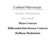
Light microscopy: topics Properties of light Compound microscope Resolution & contrast Contrast modes in light microscopy Koehler Illumination Class exercises
| PowerPoint PPT presentation | free to view

Angular Magnification, M. Near point: shortest distance at which eye can accommodate (250mm) ... there is no limit to the magnification that can be attained! ...
| PowerPoint PPT presentation | free to view

1. From cavemen to Newton: earliest ideas about light. 2. Particles and Waves: what is wave-particle duality? ... 3. Blue Skies, Rainbows and Thunder Storms: ...
| PowerPoint PPT presentation | free to download

To illustrate the parts of the microscope and their functions. ... Anatomy of a planarian. Electron Microscopes. I. Characteristics ...
| PowerPoint PPT presentation | free to view
Scanning Tunneling Microscopy (STM) - History ... constant height mode, conductance mapping, tunneling spectroscopy - Examples of STM studies ...
| PowerPoint PPT presentation | free to download

Malachite [light green/Cu2(CO3)(OH)2 & Azurite [blue/Cu3(CO3)2(OH)2. In October of 1904 the strangled body of Eva Disch was found near Frankfurt, Germany ...
| PowerPoint PPT presentation | free to view

Non-specifically stains carbohydrates. Used to demonstrate glycogen,mucin, etc. ... HIO4= Controls? Stem stained with toluidine blue. Quantitation using flow cytometry ...
| PowerPoint PPT presentation | free to view

Magnification and Orientation II. object. focal point ... Final magnification using a simple lens system (e.g., dissecting microscope. Same images: ...
| PowerPoint PPT presentation | free to view

How to Use a Compound Microscope Basic Microscopy Image: The Far Side by Gary Larson From the Virtual Microbiology Classroom on ScienceProfOnline.com
| PowerPoint PPT presentation | free to view

Light Microscopy and Electronic Imaging for the Biomedical Sciences E. D. Salmon and Kerry Bloom Biology 188 History of the Microscope, Thomas E. Jones Janssen ...
| PowerPoint PPT presentation | free to download

CZECH TECHNICAL UNIVERSITY IN PRAGUE FACULTY OF BIOMEDICAL ENGINEERING Fluorescence microscopy I Basic concepts of optical microscopy Martin Hof, Radek Mach
| PowerPoint PPT presentation | free to view

Strategy for Working up in Magnification. Focused image at 40x target circled ... 'Dry' magnification ... Focus at high dry magnification. Move the high dry ...
| PowerPoint PPT presentation | free to view

Surface plasmons represent collective excitations of electrons in metals. ... is selected as the member of Phi Beta Kappa, and one of the 2006 Nanoscience ...
| PowerPoint PPT presentation | free to download

Electron Microscopy (EM) Grid and Carbon film Preparation for Cryo-Electron Microscopy Cryo-EM tutorial July25~29, 2005 Zongli Li Some basic topics about EM grid Home ...
| PowerPoint PPT presentation | free to view

Drawings of protozoa from early work with the light microscope ... Atlas by Don Fawcett is particularly impressive. Check out some of the old papers ...
| PowerPoint PPT presentation | free to view

Darkfield Microscopy. Image is made up of highly diffracted light. Low light, can detect detail ... Phase contrast microscopy makes these phase difference visible ...
| PowerPoint PPT presentation | free to view

Staining Procedures ... Differential Stains Stain one group of organisms/cells different than another Gram stain Acid Fast Stain Microscopy Staining Techniques ...
| PowerPoint PPT presentation | free to view

In lab, use a compound microscope-multiple magnifying lenses ... of light microscope examine live ... Electron Microscope. Two types of Electron Microscopes ...
| PowerPoint PPT presentation | free to view
MICROSCOPY AND STAINING CHAPTER 3 Metric Units Light Properties Wavelength polarity Waves can be added Light Properties Resolution Wavelength/Resolution Interaction ...
| PowerPoint PPT presentation | free to download


Microscopy Lab 3 Microscopy Microscopes are used to view things that are too small to see without help. Dissecting microscope view entire 3-D objects under low power.
| PowerPoint PPT presentation | free to download

Lecture 6: Microscopy II PHYS 430/603 material Laszlo Takacs UMBC Department of Physics Light microscopy The principle and a commercial scope Optical microscope (OM ...
| PowerPoint PPT presentation | free to download

Lecture 5: Microscopy PHYS 430/603 material Laszlo Takacs UMBC Department of Physics Light microscopy The principle and a commercial scope Optical microscope (OM) and ...
| PowerPoint PPT presentation | free to view

Look in the center, dude! Chromatic aberration. Prism effect. ... You should have a total of 6 tubes: 2 deeps, 2 slants and 2 broths. ...
| PowerPoint PPT presentation | free to view

... agent for tuberculosis) easy to recognize in sputum, amidst numerous organisms. sputum sample containing. M. tuberculosis. Special Stains ...
| PowerPoint PPT presentation | free to view

Confocal Microscopy
| PowerPoint PPT presentation | free to view

Field of view vs. Depth of Field ... Depth of field-the amount of the field of view that is in sharp focus. Crossed Threads Slide ...
| PowerPoint PPT presentation | free to view
Microscopy Compound Microscope ... Endospore stain An endospore is a special structure made by some bacteria to store genetic material for a new cell.
| PowerPoint PPT presentation | free to download

Optical Microscopy Lecture 1 Some Conventions S is distance from the object; S is distance from the image Sign conventions: m = positive for inverted image ...
| PowerPoint PPT presentation | free to view
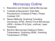
Microscopy Outline. Resolution and Simple Optical Microscope. Contrast enhancement: Dark field, Fluorescence (Chelsea & Peter), Phase Contrast, DIC
| PowerPoint PPT presentation | free to download

Transformers: 20-100 kV; Vacuum pumps: 10-6 10-10 Torr. OU NanoLab/NSF NUE/Bumm & Johnson ... TEM IMAGES. www.udel.edu ; www.nano-lab. com ; www.thermo.com ...
| PowerPoint PPT presentation | free to download
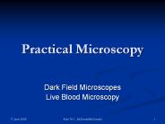
Practical Microscopy Dark Field Microscopes Live Blood Microscopy Microscopes & TH1 Disease Wirotsko, Wright, Cantwell, Mattman all published images of microbes in ...
| PowerPoint PPT presentation | free to download

di = distance from the pinhole. to the image. do = distance from the pinhole. to ... 4. Position: di = do. FLAT MIRRORS ? ... 1/do 1/di = 2/R. CURVED MIRRORS ...
| PowerPoint PPT presentation | free to view

... lenses in a tube, leading to the forerunner of the microscope and the telescope. In the late 1600's, Anton van Leeuwenhoek was the first to see bacteria, yeast, ...
| PowerPoint PPT presentation | free to view
Title: PowerPoint Presentation - Intro to Optics Author: Michael A. Rea Last modified by: Michael Rea Created Date: 3/19/2001 5:08:04 PM Document presentation format
| PowerPoint PPT presentation | free to download

hologram. Hologram reconstruction. Digital reconstruction. Numerical simulation of reference. wave diffraction by the hologram: 1) intensity contrast images ...
| PowerPoint PPT presentation | free to view

Polarized and Differential Interference Contrast (DIC) Microscopy The term polarized light refers to the properties of light waves in relation to the plane in ...
| PowerPoint PPT presentation | free to view
Title: 9 - Visualisation des cellules Subject: IA - Microscopie photonique Author: Bertrand MAC Description: Photos de MBoC4 Last modified by: CHU de Rouen
| PowerPoint PPT presentation | free to download

Permits optical sectioning by decreasing depth of field. ... In biological samples, the high turbidity makes use of a conventional microscope very difficult ...
| PowerPoint PPT presentation | free to view

Introduction to Microscopy: The Light Microscope The TEM The SEM Introduction to Microscopy (Use of Microscopes to study minute [small] objects) The image to the ...
| PowerPoint PPT presentation | free to view

Employ simple, non plan lenses to minimize. internal elements. ... Use High Quantum Efficiency Detector in Camera. Adapted from E.D.Salmon. Live Cell Considerations ...
| PowerPoint PPT presentation | free to view
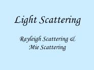
Refractive index is a weak function of wavelength. How does light interact with objects? ... Shine light source though solutions in a dark room ...
| PowerPoint PPT presentation | free to download

Title: Optical Microscopy Author: Greg Druschel Last modified by: X Created Date: 2/22/2004 9:37:42 PM Document presentation format: On-screen Show
| PowerPoint PPT presentation | free to download
Atomic Force Microscopy Lecture 7 Outline 1. Introduction to Atomic Forces 2. AFM Modes of operation 3. Case study 11: Information from AFM
| PowerPoint PPT presentation | free to download

Quantitative Fluorescence Microscopy Ana Gonz lez Wusener Instituto de Investigaciones Biotecnol gicas IIB-INTECH Universidad Nacional de General San Mart n
| PowerPoint PPT presentation | free to view

... Prefix abbreviation factor of base unit giga G 1,000,000,000 mega M 1,000,000 kilo k 1,000 hecto h 100 deka da 10 Prefix abbreviation factor of base unit deci d 0 ...
| PowerPoint PPT presentation | free to view

http://www.ispub.com/ostia/index.php?xmlPrinter=true&xmlFilePath=journals/i jmb ... http://www.phy.cuhk.edu.hk/centrallaboratory/CM120/CM120.jpg ...
| PowerPoint PPT presentation | free to view
History of the Microscope Development of the microscope ... later in life to help the progress of microbiology We measure ... Historical Microscopy 2 ...
| PowerPoint PPT presentation | free to download

Optical Microscopy. Optical or light microscopy involves passing visible light ... Confocal laser scanning microscopy (CLSM) is a relatively new light ...
| PowerPoint PPT presentation | free to view

... eye using a tool called a microscope an instrument that gives an enlarged image of the object under study Compound microscope Electron microscope Scanning ...
| PowerPoint PPT presentation | free to download

Properties of Light What is light? It is a small part of the EM spectrum, but it is the one we are most familiar with. How fast does light travel?
| PowerPoint PPT presentation | free to download

See picture Optical Microscopy Using an optical microscope in dark field ... OU NanoLab/NSF NUE/Bumm & Johnson Dark-Field Optical Microscopy * www ...
| PowerPoint PPT presentation | free to download

Most of the descriptions of the cell in your textbook are based on ... Rheostat: adjusts light intensity. Iris diaphragm (adjusts light coming in) OBJECTIVES ...
| PowerPoint PPT presentation | free to view
Basic Electron Microscopy Arthur Rowe The Knowledge Base at a Simple Level
| PowerPoint PPT presentation | free to download
A few words about microscopy Magnification Resolution Contrast Two- and three-dimensional imaging Compound light microscope Inexpensive and easy to use Uses visible ...
| PowerPoint PPT presentation | free to download